1WW2

Crystallographic studies on two bioisosteric analogues, N-acetyl-beta-D-glucopyranosylamine and N-trifluoroacetyl-beta-D-glucopyranosylamine, potent inhibitors of muscle glycogen phosphorylase


Experimental Data Snapshot

  • Method: X-RAY DIFFRACTION
  • Resolution: 1.90 Å
  • R-Value Free: 0.213 
  • R-Value Work: 0.189 
  • R-Value Observed: 0.189 

wwPDB Validation   3D Report Full Report


This is version 1.4 of the entry. See complete history


Literature

Crystallographic studies on two bioisosteric analogues, N-acetyl-beta-d-glucopyranosylamine and N-trifluoroacetyl-beta-d-glucopyranosylamine, potent inhibitors of muscle glycogen phosphorylase

Anagnostou, E.Kosmopoulou, M.N.Chrysina, E.D.Leonidas, D.D.Hadjiloi, T.Tiraidis, C.Zographos, S.E.Gyorgydeak, Z.Somsak, L.Docsa, T.Gergely, P.Kolisis, F.N.Oikonomakos, N.G.

(2006) Bioorg Med Chem 14: 181-189

  • DOI: https://doi.org/10.1016/j.bmc.2005.08.010
  • Primary Citation of Related Structures:  
    1WW2, 1WW3

  • PubMed Abstract: 

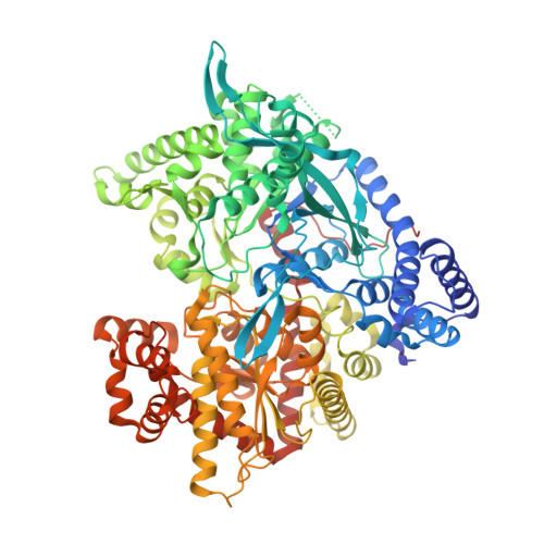
    Structure-based inhibitor design has led to the discovery of a number of potent inhibitors of glycogen phosphorylase b (GPb), N-acyl derivatives of beta-D-glucopyranosylamine, that bind at the catalytic site of the enzyme. The first good inhibitor in this class of compounds, N-acetyl-beta-D-glucopyranosylamine (NAG) (K(i) = 32 microM), has been previously characterized by biochemical, biological and crystallographic experiments at 2.3 angstroms resolution. Bioisosteric replacement of the acetyl group by trifluoroacetyl group resulted in an inhibitor, N-trifluoroacetyl-beta-D-glucopyranosylamine (NFAG), with a K(i) = 75 microM. To elucidate the structural basis of its reduced potency, we determined the ligand structure in complex with GPb at 1.8 angstroms resolution. To compare the binding mode of N-trifluoroacetyl derivative with that of the lead molecule, we also determined the structure of GPb-NAG complex at a higher resolution (1.9 angstroms). NFAG can be accommodated in the catalytic site of T-state GPb at approximately the same position as that of NAG and stabilize the T-state conformation of the 280 s loop by making several favourable contacts to Asn284 of this loop. The difference observed in the K(i) values of the two analogues can be interpreted in terms of subtle conformational changes of protein residues and shifts of water molecules in the vicinity of the catalytic site, variations in van der Waals interaction, and desolvation effects.


  • Organizational Affiliation

    Institute of Organic and Pharmaceutical Chemistry, The National Hellenic Research Foundation, 48, Vas. Constantinou Ave. 116 35 Athens, Greece.


Macromolecules
Find similar proteins by:  (by identity cutoff)  |  3D Structure
Entity ID: 1
MoleculeChains Sequence LengthOrganismDetailsImage
Glycogen phosphorylase, muscle form842Oryctolagus cuniculusMutation(s): 0 
EC: 2.4.1.1
UniProt
Find proteins for P00489 (Oryctolagus cuniculus)
Explore P00489 
Go to UniProtKB:  P00489
Entity Groups  
Sequence Clusters30% Identity50% Identity70% Identity90% Identity95% Identity100% Identity
UniProt GroupP00489
Sequence Annotations
Expand
  • Reference Sequence
Small Molecules
Ligands 2 Unique
IDChains Name / Formula / InChI Key2D Diagram3D Interactions
PLP
Query on PLP

Download Ideal Coordinates CCD File 
C [auth A]PYRIDOXAL-5'-PHOSPHATE
C8 H10 N O6 P
NGVDGCNFYWLIFO-UHFFFAOYSA-N
NBG
Query on NBG

Download Ideal Coordinates CCD File 
B [auth A]N-acetyl-beta-D-glucopyranosylamine
C8 H15 N O6
IBONACLSSOLHFU-JAJWTYFOSA-N
Binding Affinity Annotations 
IDSourceBinding Affinity
NBG BindingDB:  1WW2 Ki: min: 3.16e+4, max: 3.20e+4 (nM) from 2 assay(s)
Binding MOAD:  1WW2 Ki: 3.20e+4 (nM) from 1 assay(s)
Experimental Data & Validation

Experimental Data

  • Method: X-RAY DIFFRACTION
  • Resolution: 1.90 Å
  • R-Value Free: 0.213 
  • R-Value Work: 0.189 
  • R-Value Observed: 0.189 
  • Space Group: P 43 21 2
Unit Cell:
Length ( Å )Angle ( ˚ )
a = 128.752α = 90
b = 128.752β = 90
c = 116.121γ = 90
Software Package:
Software NamePurpose
CNSrefinement
HKL-2000data reduction
SCALEPACKdata scaling
CNSphasing

Structure Validation

View Full Validation Report


Entry History 

Revision History  (Full details and data files)

  • Version 1.0: 2005-12-13
    Type: Initial release
  • Version 1.1: 2008-04-30
    Changes: Version format compliance
  • Version 1.2: 2011-07-13
    Changes: Derived calculations, Version format compliance
  • Version 1.3: 2020-07-29
    Type: Remediation
    Reason: Carbohydrate remediation
    Changes: Data collection, Database references, Derived calculations, Structure summary
  • Version 1.4: 2023-10-25
    Changes: Data collection, Database references, Refinement description, Structure summary