Crystal Structures of Biotin Protein Ligase from Pyrococcus horikoshii OT3 and its Complexes: Structural Basis of Biotin Activation
Bagautdinov, B., Kuroishi, C., Sugahara, M., Kunishima, N.(2005) J Mol Biology 353: 322-333
- PubMed: 16169557 Search on PubMed
- DOI: https://doi.org/10.1016/j.jmb.2005.08.032
- Primary Citation Related Structures:
1WNL, 1WPY, 1WQ7, 1WQW - PubMed Abstract:
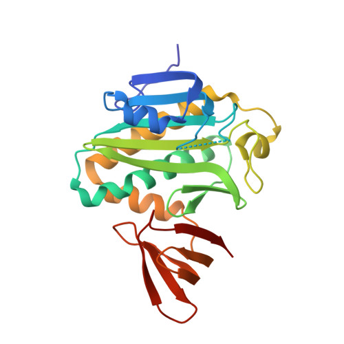
Biotin protein ligase (EC 6.3.4.15) catalyses the synthesis of an activated form of biotin, biotinyl-5'-AMP, from substrates biotin and ATP followed by biotinylation of the biotin carboxyl carrier protein subunit of acetyl-CoA carboxylase. The three-dimensional structure of biotin protein ligase from Pyrococcus horikoshii OT3 has been determined by X-ray diffraction at 1.6A resolution. The structure reveals a homodimer as the functional unit. Each subunit contains two domains, a larger N-terminal catalytic domain and a smaller C-terminal domain. The structural feature of the active site has been studied by determination of the crystal structures of complexes of the enzyme with biotin, ADP and the reaction intermediate biotinyl-5'-AMP at atomic resolution. This is the first report of the liganded structures of biotin protein ligase with nucleotide and biotinyl-5'-AMP. The structures of the unliganded and the liganded forms are isomorphous except for an ordering of the active site loop upon ligand binding. Catalytic binding sites are suitably arranged to minimize the conformational changes required during the reaction, as the pockets for biotin and nucleotide are located spatially adjacent to each other in a cleft of the catalytic domain and the pocket for biotinyl-5'-AMP binding mimics the combination of those of the substrates. The exact locations of the ligands and the active site residues allow us to propose a general scheme for the first step of the reaction carried out by biotin protein ligase in which the positively charged epsilon-amino group of Lys111 facilitates the nucleophilic attack on the ATP alpha-phosphate group by the biotin carboxyl oxygen atom and stabilizes the negatively charged intermediates.
- Advanced Protein Crystallography Research Group, RIKEN Harima Institute at SPring-8, 1-1-1 Kouto, Mikazuki-cho, Sayo-gun, Hyogo 679-5148, Japan.
Organizational Affiliation: