1VCL

Crystal Structure of Hemolytic Lectin CEL-III


Experimental Data Snapshot

  • Method: X-RAY DIFFRACTION
  • Resolution: 1.70 Å
  • R-Value Free: 0.201 
  • R-Value Work: 0.165 
  • R-Value Observed: 0.167 

wwPDB Validation   3D Report Full Report


This is version 2.1 of the entry. See complete history


Literature

Crystal Structure of the Hemolytic Lectin CEL-III Isolated from the Marine Invertebrate Cucumaria echinata: IMPLICATIONS OF DOMAIN STRUCTURE FOR ITS MEMBRANE PORE-FORMATION MECHANISM

Uchida, T.Yamasaki, T.Eto, S.Sugawara, H.Kurisu, G.Nakagawa, A.Kusunoki, M.Hatakeyama, T.

(2004) J Biol Chem 279: 37133-37141

  • DOI: https://doi.org/10.1074/jbc.M404065200
  • Primary Citation of Related Structures:  
    1VCL

  • PubMed Abstract: 

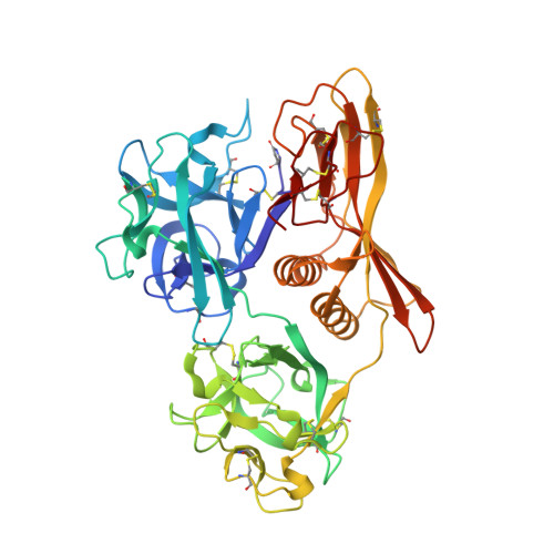
    CEL-III is a Ca(2+)-dependent and galactose-specific lectin purified from the sea cucumber, Cucumaria echinata, which exhibits hemolytic and hemagglutinating activities. Six molecules of CEL-III are assumed to oligomerize to form an ion-permeable pore in the cell membrane. We have determined the crystal structure of CELIII by using single isomorphous replacement aided by anomalous scattering in lead at 1.7 A resolution. CEL-III consists of three distinct domains as follows: the N-terminal two carbohydrate-binding domains (1 and 2), which adopt beta-trefoil folds such as the B-chain of ricin and are members of the (QXW)(3) motif family; and domain 3, which is a novel fold composed of two alpha-helices and one beta-sandwich. CEL-III is the first Ca(2+)-dependent lectin structure with two beta-trefoil folds. Despite sharing the structure of the B-chain of ricin, CEL-III binds five Ca(2+) ions at five of the six subdomains in both domains 1 and 2. Considering the relatively high similarity among the five subdomains, they are putative binding sites for galactose-related carbohydrates, although it remains to be elucidated whether bound Ca(2+) is directly involved in interaction with carbohydrates. The paucity of hydrophobic interactions in the interfaces between the domains and biochemical data suggest that these domains rearrange upon carbohydrate binding in the erythrocyte membrane. This conformational change may be responsible for oligomerization of CEL-III molecules and hemolysis in the erythrocyte membranes.


  • Organizational Affiliation

    Research Center for Structural and Functional Proteomics, Institute for Protein Research, Osaka University, 3-2 Yamadaoka, Suita, Osaka 565-0871.


Macromolecules
Find similar proteins by:  (by identity cutoff)  |  3D Structure
Entity ID: 1
MoleculeChains Sequence LengthOrganismDetailsImage
hemolytic lectin CEL-III
A, B
432Cucumaria echinataMutation(s): 0 
UniProt
Find proteins for Q868M7 (Cucumaria echinata)
Explore Q868M7 
Go to UniProtKB:  Q868M7
Entity Groups  
Sequence Clusters30% Identity50% Identity70% Identity90% Identity95% Identity100% Identity
UniProt GroupQ868M7
Sequence Annotations
Expand
  • Reference Sequence
Small Molecules
Ligands 4 Unique
IDChains Name / Formula / InChI Key2D Diagram3D Interactions
BTB
Query on BTB

Download Ideal Coordinates CCD File 
K [auth A]2-[BIS-(2-HYDROXY-ETHYL)-AMINO]-2-HYDROXYMETHYL-PROPANE-1,3-DIOL
C8 H19 N O5
OWMVSZAMULFTJU-UHFFFAOYSA-N
CA
Query on CA

Download Ideal Coordinates CCD File 
C [auth A]
D [auth A]
E [auth A]
F [auth A]
G [auth A]
C [auth A],
D [auth A],
E [auth A],
F [auth A],
G [auth A],
L [auth B],
M [auth B],
N [auth B],
O [auth B],
P [auth B]
CALCIUM ION
Ca
BHPQYMZQTOCNFJ-UHFFFAOYSA-N
CL
Query on CL

Download Ideal Coordinates CCD File 
J [auth A],
S [auth B]
CHLORIDE ION
Cl
VEXZGXHMUGYJMC-UHFFFAOYSA-M
MG
Query on MG

Download Ideal Coordinates CCD File 
H [auth A],
I [auth A],
Q [auth B],
R [auth B]
MAGNESIUM ION
Mg
JLVVSXFLKOJNIY-UHFFFAOYSA-N
Modified Residues  1 Unique
IDChains TypeFormula2D DiagramParent
PCA
Query on PCA
A, B
L-PEPTIDE LINKINGC5 H7 N O3GLN
Experimental Data & Validation

Experimental Data

  • Method: X-RAY DIFFRACTION
  • Resolution: 1.70 Å
  • R-Value Free: 0.201 
  • R-Value Work: 0.165 
  • R-Value Observed: 0.167 
  • Space Group: P 1 21 1
Unit Cell:
Length ( Å )Angle ( ˚ )
a = 52.42α = 90
b = 65.37β = 98.18
c = 126.02γ = 90
Software Package:
Software NamePurpose
REFMACrefinement
MOSFLMdata reduction
CCP4data scaling
SHARPphasing

Structure Validation

View Full Validation Report


Entry History 

Deposition Data

Revision History  (Full details and data files)

  • Version 1.0: 2004-09-07
    Type: Initial release
  • Version 1.1: 2008-04-27
    Changes: Version format compliance
  • Version 1.2: 2011-07-13
    Changes: Version format compliance
  • Version 2.0: 2019-12-25
    Changes: Database references, Derived calculations, Polymer sequence
  • Version 2.1: 2023-12-27
    Changes: Data collection, Database references, Derived calculations