1UKZ

SUBSTRATE SPECIFICITY AND ASSEMBLY OF CATALYTIC CENTER DERIVED FROM TWO STRUCTURES OF LIGATED URIDYLATE KINASE


Experimental Data Snapshot

  • Method: X-RAY DIFFRACTION
  • Resolution: 1.90 Å
  • R-Value Work: 0.196 
  • R-Value Observed: 0.196 

wwPDB Validation   3D Report Full Report


Ligand Structure Quality Assessment 


This is version 1.2 of the entry. See complete history


Literature

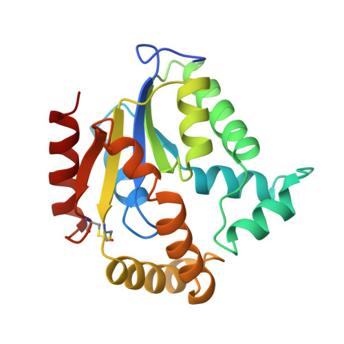
Substrate specificity and assembly of the catalytic center derived from two structures of ligated uridylate kinase.

Muller-Dieckmann, H.J.Schulz, G.E.

(1995) J Mol Biol 246: 522-530

  • DOI: https://doi.org/10.1006/jmbi.1994.0104
  • Primary Citation of Related Structures:  
    1UKY, 1UKZ

  • PubMed Abstract: 

    Two crystal structures of ligated uridylate kinase from Saccharomyces cerevisiae were determined by X-ray analyses. The ligands were ADP and AMP. Cocrystallization with ATP yielded crystals with ADP at the ATP site and a mixture of AMP and ADP at the NMP site. Cocrystallization with ADP gave rise to a distinct crystal type with ADP at the ATP site, but only AMP at the NMP site. In both cases, the substrates are kept in place by favorable crystal contacts. The structures have been refined to R-factors of 17.8% and 19.6% at resolutions of 2.1 A and 1.9 A, respectively. A comparison with the related cytosolic adenylate kinase from pig disclosed large induced-fit movements on substrate binding and the disassembly of the catalytic center in the absence of substrates. The relatively high side-activity of uridylate kinase for AMP is explained by the finding that the binding pocket is sized for an AMP, but constructed to bind UMP together with a water molecule.


  • Organizational Affiliation

    Institut für Organische, Chemie und Biochemie, Albert-Ludwigs-Universität, Freiburg im Breisgau, Germany.


Macromolecules
Find similar proteins by:  (by identity cutoff)  |  3D Structure
Entity ID: 1
MoleculeChains Sequence LengthOrganismDetailsImage
URIDYLATE KINASE203Saccharomyces cerevisiaeMutation(s): 0 
EC: 2.7.4
UniProt
Find proteins for P15700 (Saccharomyces cerevisiae (strain ATCC 204508 / S288c))
Explore P15700 
Go to UniProtKB:  P15700
Entity Groups  
Sequence Clusters30% Identity50% Identity70% Identity90% Identity95% Identity100% Identity
UniProt GroupP15700
Sequence Annotations
Expand
  • Reference Sequence
Small Molecules
Ligands 2 Unique
IDChains Name / Formula / InChI Key2D Diagram3D Interactions
ADP
Query on ADP

Download Ideal Coordinates CCD File 
B [auth A]ADENOSINE-5'-DIPHOSPHATE
C10 H15 N5 O10 P2
XTWYTFMLZFPYCI-KQYNXXCUSA-N
AMP
Query on AMP

Download Ideal Coordinates CCD File 
C [auth A]ADENOSINE MONOPHOSPHATE
C10 H14 N5 O7 P
UDMBCSSLTHHNCD-KQYNXXCUSA-N
Experimental Data & Validation

Experimental Data

  • Method: X-RAY DIFFRACTION
  • Resolution: 1.90 Å
  • R-Value Work: 0.196 
  • R-Value Observed: 0.196 
  • Space Group: P 61 2 2
Unit Cell:
Length ( Å )Angle ( ˚ )
a = 63.34α = 90
b = 63.34β = 90
c = 183.35γ = 120
Software Package:
Software NamePurpose
X-PLORmodel building
X-PLORrefinement
X-PLORphasing

Structure Validation

View Full Validation Report


Ligand Structure Quality Assessment 


Entry History 

Deposition Data

Revision History  (Full details and data files)

  • Version 1.0: 1995-01-26
    Type: Initial release
  • Version 1.1: 2008-03-24
    Changes: Version format compliance
  • Version 1.2: 2011-07-13
    Changes: Version format compliance