1UGD

HUMAN CARBONIC ANHYDRASE II[HCAII] (E.C.4.2.1.1) MUTANT WITH ALA 65 REPLACED BY SER (A65S)


Experimental Data Snapshot

  • Method: X-RAY DIFFRACTION
  • Resolution: 2.00 Å
  • R-Value Free: 0.255 
  • R-Value Work: 0.176 
  • R-Value Observed: 0.176 

wwPDB Validation   3D Report Full Report


This is version 1.4 of the entry. See complete history


Literature

X-ray crystallographic studies of alanine-65 variants of carbonic anhydrase II reveal the structural basis of compromised proton transfer in catalysis.

Scolnick, L.R.Christianson, D.W.

(1996) Biochemistry 35: 16429-16434

  • DOI: https://doi.org/10.1021/bi9617872
  • Primary Citation of Related Structures:  
    1UGA, 1UGB, 1UGC, 1UGD, 1UGE, 1UGF, 1UGG

  • PubMed Abstract: 

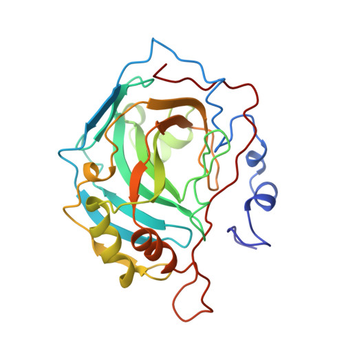
    The three-dimensional structures of A65F, A65L, A65H, A65T, A65S, and A65G human carbonic anhydrase II (CAII) variants have been solved by X-ray crystallographic methods to probe the importance of residue 65 and the structural implications of its evolutionary drift in the greater family of carbonic anhydrase isozymes. Structure-activity relationships in this series of CAII variants are correlated with those established for other carbonic anhydrase isozymes. We conclude that a bulky side chain at position 65 hinders the formation of an effective solvent bridge between zinc-bound water and H64 and thereby hinders solvent-mediated proton transfer between these two groups [Jackman, J. E., Merz, K. M., Jr., & Fierke, C. A. (1996) Biochemistry 35, 16421-16428]. Despite the introduction of a polar hydroxyl group at this position, smaller side chains such as serine or threonine substituted for A65 do not perturb the formation of a solvent bridge between H64 and zinc-bound solvent. Thus, the evolution of residue 65 size is one factor affecting the trajectory of catalytic proton transfer.


  • Organizational Affiliation

    Department of Chemistry, University of Pennsylvania, Philadelphia 19104-6323, USA.


Macromolecules
Find similar proteins by:  (by identity cutoff)  |  3D Structure
Entity ID: 1
MoleculeChains Sequence LengthOrganismDetailsImage
CARBONIC ANHYDRASE II258Homo sapiensMutation(s): 1 
Gene Names: CAII
EC: 4.2.1.1
UniProt & NIH Common Fund Data Resources
Find proteins for P00918 (Homo sapiens)
Explore P00918 
Go to UniProtKB:  P00918
PHAROS:  P00918
GTEx:  ENSG00000104267 
Entity Groups  
Sequence Clusters30% Identity50% Identity70% Identity90% Identity95% Identity100% Identity
UniProt GroupP00918
Sequence Annotations
Expand
  • Reference Sequence
Small Molecules
Ligands 1 Unique
IDChains Name / Formula / InChI Key2D Diagram3D Interactions
ZN
Query on ZN

Download Ideal Coordinates CCD File 
B [auth A]ZINC ION
Zn
PTFCDOFLOPIGGS-UHFFFAOYSA-N
Experimental Data & Validation

Experimental Data

  • Method: X-RAY DIFFRACTION
  • Resolution: 2.00 Å
  • R-Value Free: 0.255 
  • R-Value Work: 0.176 
  • R-Value Observed: 0.176 
  • Space Group: P 1 21 1
Unit Cell:
Length ( Å )Angle ( ˚ )
a = 42.7α = 90
b = 41.7β = 104.6
c = 73γ = 90
Software Package:
Software NamePurpose
X-PLORmodel building
X-PLORrefinement
MOSFLMdata reduction
Agrovatadata scaling
ROTAVATAdata scaling
X-PLORphasing

Structure Validation

View Full Validation Report


Entry History 

Deposition Data

Revision History  (Full details and data files)

  • Version 1.0: 1997-01-27
    Type: Initial release
  • Version 1.1: 2008-03-24
    Changes: Version format compliance
  • Version 1.2: 2011-07-13
    Changes: Version format compliance
  • Version 1.3: 2021-11-03
    Changes: Database references, Derived calculations, Other, Refinement description
  • Version 1.4: 2024-02-14
    Changes: Data collection