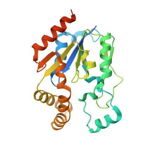
1UFQ

Crystal structure of ligand-free human uridine-cytidine kinase 2


Experimental Data Snapshot

  • Method: X-RAY DIFFRACTION
  • Resolution: 2.50 Å
  • R-Value Free: 0.287 
  • R-Value Work: 0.236 
  • R-Value Observed: 0.237 

wwPDB Validation   3D Report Full Report


This is version 1.3 of the entry. See complete history


Literature

Structural basis for the specificity, catalysis, and regulation of human uridine-cytidine kinase

Suzuki, N.N.Koizumi, K.Fukushima, M.Matsuda, A.Inagaki, F.

(2004) Structure 12: 751-764

  • DOI: https://doi.org/10.1016/j.str.2004.02.038
  • Primary Citation of Related Structures:  
    1UDW, 1UEI, 1UEJ, 1UFQ, 1UJ2

  • PubMed Abstract: 

    Uridine-cytidine kinase (UCK) catalyzes the phosphorylation of uridine and cytidine and activates pharmacological ribonucleoside analogs. Here we present the crystal structures of human UCK alone and in complexes with a substrate, cytidine, a feedback inhibitor, CTP or UTP, and with phosphorylation products, CMP and ADP, respectively. Free UCK takes an alpha/beta mononucleotide binding fold and exists as a homotetramer with 222 symmetry. Upon inhibitor binding, one loop region was loosened, causing the UCK tetramer to be distorted. Upon cytidine binding, a large induced fit was observed at the uridine/cytidine binding site, which endows UCK with a strict specificity for pyrimidine ribonucleosides. The first UCK structure provided the structural basis for the specificity, catalysis, and regulation of human uridine-cytidine kinase, which give clues for the design of novel antitumor and antiviral ribonucleoside analogs that inhibit RNA synthesis.


  • Organizational Affiliation

    Department of Structural Biology, Graduate School of Pharmaceutical Sciences, Hokkaido University, N12 W6 Kita-ku, Sapporo 060-0812, Japan.


Macromolecules
Find similar proteins by:  (by identity cutoff)  |  3D Structure
Entity ID: 1
MoleculeChains Sequence LengthOrganismDetailsImage
Uridine-cytidine kinase 2
A, B, C, D
252Homo sapiensMutation(s): 0 
EC: 2.7.1.48
UniProt & NIH Common Fund Data Resources
Find proteins for Q9BZX2 (Homo sapiens)
Explore Q9BZX2 
Go to UniProtKB:  Q9BZX2
PHAROS:  Q9BZX2
GTEx:  ENSG00000143179 
Entity Groups  
Sequence Clusters30% Identity50% Identity70% Identity90% Identity95% Identity100% Identity
UniProt GroupQ9BZX2
Sequence Annotations
Expand
  • Reference Sequence
Experimental Data & Validation

Experimental Data

  • Method: X-RAY DIFFRACTION
  • Resolution: 2.50 Å
  • R-Value Free: 0.287 
  • R-Value Work: 0.236 
  • R-Value Observed: 0.237 
  • Space Group: P 21 21 2
Unit Cell:
Length ( Å )Angle ( ˚ )
a = 84.057α = 90
b = 94.262β = 90
c = 156.989γ = 90
Software Package:
Software NamePurpose
CNSrefinement
CrystalCleardata reduction
CrystalCleardata scaling
CNSphasing

Structure Validation

View Full Validation Report


Entry History 

Deposition Data

Revision History  (Full details and data files)

  • Version 1.0: 2004-05-04
    Type: Initial release
  • Version 1.1: 2008-04-27
    Changes: Version format compliance
  • Version 1.2: 2011-07-13
    Changes: Version format compliance
  • Version 1.3: 2023-10-25
    Changes: Data collection, Database references, Refinement description