1UA1

Structure of aminofluorene adduct paired opposite cytosine at the polymerase active site.


Experimental Data Snapshot

  • Method: X-RAY DIFFRACTION
  • Resolution: 2.00 Å
  • R-Value Free: 0.249 
  • R-Value Work: 0.216 
  • R-Value Observed: 0.216 

wwPDB Validation   3D Report Full Report


Ligand Structure Quality Assessment 


This is version 1.3 of the entry. See complete history


Literature

Observing translesion synthesis of an aromatic amine DNA adduct by a high-fidelity DNA polymerase

Hsu, G.W.Kiefer, J.R.Becherel, O.J.Fuchs, R.P.P.Beese, L.S.

(2004) J Biol Chem 279: 50280-50285

  • DOI: https://doi.org/10.1074/jbc.M409224200
  • Primary Citation of Related Structures:  
    1UA1

  • PubMed Abstract: 

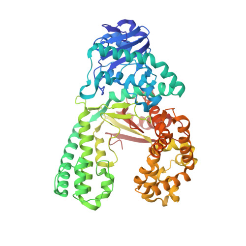
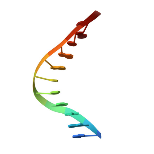
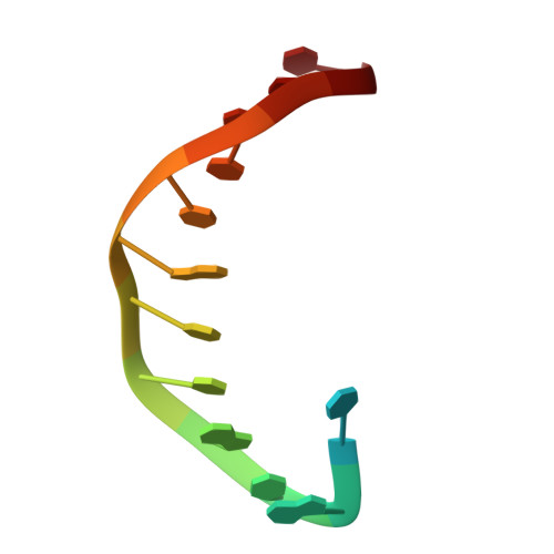
    Aromatic amines have been studied for more than a half-century as model carcinogens representing a class of chemicals that form bulky adducts to the C8 position of guanine in DNA. Among these guanine adducts, the N-(2'-deoxyguanosin-8-yl)-aminofluorene (G-AF) and N-2-(2'-deoxyguanosin-8-yl)-acetylaminofluorene (G-AAF) derivatives are the best studied. Although G-AF and G-AAF differ by only an acetyl group, they exert different effects on DNA replication by replicative and high-fidelity DNA polymerases. Translesion synthesis of G-AF is achieved with high-fidelity polymerases, whereas replication of G-AAF requires specialized bypass polymerases. Here we have presented structures of G-AF as it undergoes one round of accurate replication by a high-fidelity DNA polymerase. Nucleotide incorporation opposite G-AF is achieved in solution and in the crystal, revealing how the polymerase accommodates and replicates past G-AF, but not G-AAF. Like an unmodified guanine, G-AF adopts a conformation that allows it to form Watson-Crick hydrogen bonds with an opposing cytosine that results in protrusion of the bulky fluorene moiety into the major groove. Although incorporation opposite G-AF is observed, the C:G-AF base pair induces distortions to the polymerase active site that slow translesion synthesis.


  • Organizational Affiliation

    Department of Biochemistry, Duke University Medical Center, Durham, North Carolina 27710, USA.


Macromolecules

Find similar proteins by:  (by identity cutoff)  |  3D Structure
Entity ID: 3
MoleculeChains Sequence LengthOrganismDetailsImage
DNA polymerase IC [auth A]580Geobacillus stearothermophilusMutation(s): 0 
EC: 2.7.7.7
UniProt
Find proteins for P52026 (Geobacillus stearothermophilus)
Explore P52026 
Go to UniProtKB:  P52026
Entity Groups  
Sequence Clusters30% Identity50% Identity70% Identity90% Identity95% Identity100% Identity
UniProt GroupP52026
Sequence Annotations
Expand
  • Reference Sequence

Find similar nucleic acids by:  Sequence   |   3D Structure  

Entity ID: 1
MoleculeChains LengthOrganismImage
DNA primer strandA [auth B]11N/A
Sequence Annotations
Expand
  • Reference Sequence

Find similar nucleic acids by:  Sequence   |   3D Structure  

Entity ID: 2
MoleculeChains LengthOrganismImage
DNA template strand with aminofluorene adductB [auth C]14N/A
Sequence Annotations
Expand
  • Reference Sequence
Experimental Data & Validation

Experimental Data

  • Method: X-RAY DIFFRACTION
  • Resolution: 2.00 Å
  • R-Value Free: 0.249 
  • R-Value Work: 0.216 
  • R-Value Observed: 0.216 
  • Space Group: P 21 21 21
Unit Cell:
Length ( Å )Angle ( ˚ )
a = 87.639α = 90
b = 93.441β = 90
c = 105.465γ = 90
Software Package:
Software NamePurpose
DENZOdata reduction
SCALEPACKdata scaling
CNSrefinement
CNSphasing

Structure Validation

View Full Validation Report


Ligand Structure Quality Assessment 


Entry History 

Deposition Data

Revision History  (Full details and data files)

  • Version 1.0: 2004-09-28
    Type: Initial release
  • Version 1.1: 2008-04-30
    Changes: Version format compliance
  • Version 1.2: 2011-07-13
    Changes: Version format compliance
  • Version 1.3: 2024-02-14
    Changes: Data collection, Database references, Derived calculations