1U87

Crystal Structure Of The 26 Kda Glutathione S-Transferase Y7F mutant From Schistosoma Japonicum Complexed With Glutathione


Experimental Data Snapshot

  • Method: X-RAY DIFFRACTION
  • Resolution: 3.50 Å
  • R-Value Free: 0.271 
  • R-Value Work: 0.199 
  • R-Value Observed: 0.199 

Starting Model: experimental
View more details

wwPDB Validation   3D Report Full Report


Ligand Structure Quality Assessment 


This is version 1.6 of the entry. See complete history


Literature

Crystallographic and Thermodynamic Analysis of the Binding of S-Octylglutathione to the Tyr 7 to Phe Mutant of Glutathione S-Transferase from Schistosoma japonicum(,)

Andujar-Sanchez, M.Smith, A.W.Clemente-Jimenez, J.M.Rodriguez-Vico, F.Las Heras-Vazquez, F.J.Jara-Perez, V.Camara-Artigas, A.

(2005) Biochemistry 44: 1174-1183

  • DOI: https://doi.org/10.1021/bi0483110
  • Primary Citation of Related Structures:  
    1U87, 1U88

  • PubMed Abstract: 

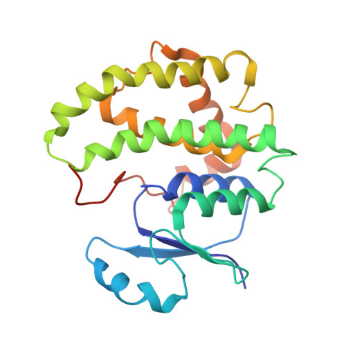
    Glutathione S-transferases are a family of multifunctional enzymes involved in the metabolism of drugs and xenobiotics. Two tyrosine residues, Tyr 7 and Tyr 111, in the active site of the enzyme play an important role in the binding and catalysis of substrate ligands. The crystal structures of Schistosoma japonicum glutathione S-transferase tyrosine 7 to phenylalanine mutant [SjGST(Y7F)] in complex with the substrate glutathione (GSH) and the competitive inhibitor S-octylglutathione (S-octyl-GSH) have been obtained. These new structural data combined with fluorescence spectroscopy and thermodynamic data, obtained by means of isothermal titration calorimetry, allow for detailed characterization of the ligand-binding process. The binding of S-octyl-GSH to SjGST(Y7F) is enthalpically and entropically driven at temperatures below 30 degrees C. The stoichiometry of the binding is one molecule of S-octyl-GSH per mutant dimer, whereas shorter alkyl derivatives bind with a stoichiometry of two molecules per mutant dimer. The SjGST(Y7F).GSH structure showed no major structural differences compared to the wild-type enzyme. In contrast, the structure of SjGST(Y7F).S-octyl-GSH showed asymmetric binding of S-octyl-GSH. This lack of symmetry is reflected in the lower symmetry space group of the SjGST(Y7F).S-octyl-GSH crystals (P6(3)) compared to that of the SjGST(Y7F).GSH crystals (P6(3)22). Moreover, the binding of S-octyl-GSH to the A subunit is accompanied by conformational changes that may be responsible for the lack of binding to the B subunit.


  • Organizational Affiliation

    Departamento Química Física, Bioquímica y Química Inorgánica, Universidad de Almería, España.


Macromolecules
Find similar proteins by:  (by identity cutoff)  |  3D Structure
Entity ID: 1
MoleculeChains Sequence LengthOrganismDetailsImage
Glutathione S-Transferase 26 kDa218Schistosoma japonicumMutation(s): 1 
EC: 2.5.1.18
UniProt
Find proteins for P08515 (Schistosoma japonicum)
Explore P08515 
Go to UniProtKB:  P08515
Entity Groups  
Sequence Clusters30% Identity50% Identity70% Identity90% Identity95% Identity100% Identity
UniProt GroupP08515
Sequence Annotations
Expand
  • Reference Sequence
Small Molecules
Ligands 1 Unique
IDChains Name / Formula / InChI Key2D Diagram3D Interactions
GSH
Query on GSH

Download Ideal Coordinates CCD File 
B [auth A]GLUTATHIONE
C10 H17 N3 O6 S
RWSXRVCMGQZWBV-WDSKDSINSA-N
Experimental Data & Validation

Experimental Data

  • Method: X-RAY DIFFRACTION
  • Resolution: 3.50 Å
  • R-Value Free: 0.271 
  • R-Value Work: 0.199 
  • R-Value Observed: 0.199 
  • Space Group: P 63 2 2
Unit Cell:
Length ( Å )Angle ( ˚ )
a = 123.05α = 90
b = 123.05β = 90
c = 72.51γ = 120
Software Package:
Software NamePurpose
CNSrefinement
MOSFLMdata reduction
CCP4data scaling
CNSphasing

Structure Validation

View Full Validation Report


Ligand Structure Quality Assessment 


Entry History 

Deposition Data

Revision History  (Full details and data files)

  • Version 1.0: 2005-02-22
    Type: Initial release
  • Version 1.1: 2008-04-30
    Changes: Version format compliance
  • Version 1.2: 2011-07-13
    Changes: Version format compliance
  • Version 1.3: 2012-03-21
    Changes: Non-polymer description
  • Version 1.4: 2017-10-11
    Changes: Data collection
  • Version 1.5: 2021-11-10
    Changes: Database references, Derived calculations
  • Version 1.6: 2023-10-25
    Changes: Data collection, Refinement description