1U0H

STRUCTURAL BASIS FOR THE INHIBITION OF MAMMALIAN ADENYLYL CYCLASE BY MANT-GTP


Experimental Data Snapshot

  • Method: X-RAY DIFFRACTION
  • Resolution: 2.90 Å
  • R-Value Free: 0.280 
  • R-Value Work: 0.245 

wwPDB Validation   3D Report Full Report


Ligand Structure Quality Assessment 


This is version 1.3 of the entry. See complete history


Literature

Structural basis for the inhibition of mammalian membrane adenylyl cyclase by 2 '(3')-O-(N-Methylanthraniloyl)-guanosine 5 '-triphosphate.

Mou, T.C.Gille, A.Fancy, D.A.Seifert, R.Sprang, S.R.

(2005) J Biol Chem 280: 7253-7261

  • DOI: https://doi.org/10.1074/jbc.M409076200
  • Primary Citation of Related Structures:  
    1TL7, 1U0H

  • PubMed Abstract: 

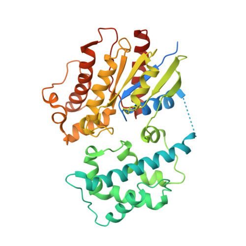
    Membrane-bound mammalian adenylyl cyclase (mAC) catalyzes the synthesis of intracellular cyclic AMP from ATP and is activated by stimulatory G protein alpha subunits (Galpha(s)) and by forskolin (FSK). mACs are inhibited with high potency by 2 '(3')-O-(N-methylanthraniloyl) (MANT)-substituted nucleotides. In this study, the crystal structures of the complex between Galpha(s).GTPgammaS and the catalytic C1 and C2 domains from type V and type II mAC (VC1.IIC2), bound to FSK and either MANT-GTP.Mg(2+) or MANT-GTP.Mn(2+) have been determined. MANT-GTP coordinates two metal ions and occupies the same position in the catalytic site as P-site inhibitors and substrate analogs. However, the orientation of the guanine ring is reversed relative to that of the adenine ring. The MANT fluorophore resides in a hydrophobic pocket at the interface between the VC1 and IIC2 domains and prevents mAC from undergoing the "open" to "closed" domain rearrangement. The K(i) of MANT-GTP for inhibition of VC1.IIC2 is lower in the presence of mAC activators and lower in the presence of Mn(2+) compared with Mg(2+), indicating that the inhibitor binds more tightly to the catalytically most active form of the enzyme. Fluorescence resonance energy transfer-stimulated emission from the MANT fluorophore upon excitation of Trp-1020 in the MANT-binding pocket of IIC2 is also stronger in the presence of FSK. Mutational analysis of two non-conserved amino acids in the MANT-binding pocket suggests that residues outside of the binding site influence isoform selectivity toward MANT-GTP.


  • Organizational Affiliation

    Department of Biochemistry, Howard Hughes Medical Institute, The University of Texas Southwestern Medical, Dallas, Texas 75390-9050, USA.


Macromolecules
Find similar proteins by:  (by identity cutoff)  |  3D Structure
Entity ID: 1
MoleculeChains Sequence LengthOrganismDetailsImage
Adenylate cyclase, type V225Canis lupus familiarisMutation(s): 0 
Gene Names: ADCY5
EC: 4.6.1.1
UniProt
Find proteins for P30803 (Canis lupus familiaris)
Explore P30803 
Go to UniProtKB:  P30803
Entity Groups  
Sequence Clusters30% Identity50% Identity70% Identity90% Identity95% Identity100% Identity
UniProt GroupP30803
Sequence Annotations
Expand
  • Reference Sequence
Find similar proteins by:  (by identity cutoff)  |  3D Structure
Entity ID: 2
MoleculeChains Sequence LengthOrganismDetailsImage
Adenylate cyclase, type II212Rattus norvegicusMutation(s): 0 
Gene Names: Adcy2
EC: 4.6.1.1
UniProt
Find proteins for P26769 (Rattus norvegicus)
Explore P26769 
Go to UniProtKB:  P26769
Entity Groups  
Sequence Clusters30% Identity50% Identity70% Identity90% Identity95% Identity100% Identity
UniProt GroupP26769
Sequence Annotations
Expand
  • Reference Sequence
Find similar proteins by:  (by identity cutoff)  |  3D Structure
Entity ID: 3
MoleculeChains Sequence LengthOrganismDetailsImage
Guanine nucleotide-binding protein G(s), alpha subunit394Bos taurusMutation(s): 0 
Gene Names: GNAS
UniProt
Find proteins for P04896 (Bos taurus)
Explore P04896 
Go to UniProtKB:  P04896
Entity Groups  
Sequence Clusters30% Identity50% Identity70% Identity90% Identity95% Identity100% Identity
UniProt GroupP04896
Sequence Annotations
Expand
  • Reference Sequence
Small Molecules
Ligands 5 Unique
IDChains Name / Formula / InChI Key2D Diagram3D Interactions
ONM
Query on ONM

Download Ideal Coordinates CCD File 
G [auth A]3'-O-(N-METHYLANTHRANILOYL)-GUANOSINE-5'-TRIPHOSPHATE
C18 H23 N6 O15 P3
DSPRYHPLXXUNHS-XNIJJKJLSA-N
GSP
Query on GSP

Download Ideal Coordinates CCD File 
J [auth C]5'-GUANOSINE-DIPHOSPHATE-MONOTHIOPHOSPHATE
C10 H16 N5 O13 P3 S
XOFLBQFBSOEHOG-UUOKFMHZSA-N
FOK
Query on FOK

Download Ideal Coordinates CCD File 
F [auth A]FORSKOLIN
C22 H34 O7
OHCQJHSOBUTRHG-KGGHGJDLSA-N
CL
Query on CL

Download Ideal Coordinates CCD File 
I [auth C]CHLORIDE ION
Cl
VEXZGXHMUGYJMC-UHFFFAOYSA-M
MG
Query on MG

Download Ideal Coordinates CCD File 
D [auth A],
E [auth A],
H [auth C]
MAGNESIUM ION
Mg
JLVVSXFLKOJNIY-UHFFFAOYSA-N
Binding Affinity Annotations 
IDSourceBinding Affinity
ONM PDBBind:  1U0H Ki: 110 (nM) from 1 assay(s)
Experimental Data & Validation

Experimental Data

  • Method: X-RAY DIFFRACTION
  • Resolution: 2.90 Å
  • R-Value Free: 0.280 
  • R-Value Work: 0.245 
  • Space Group: P 21 21 2
Unit Cell:
Length ( Å )Angle ( ˚ )
a = 118.4α = 90
b = 133β = 90
c = 70.4γ = 90
Software Package:
Software NamePurpose
CNSrefinement
HKL-2000data reduction
SCALEPACKdata scaling
CNSphasing

Structure Validation

View Full Validation Report


Ligand Structure Quality Assessment 


Entry History 

Deposition Data

Revision History  (Full details and data files)

  • Version 1.0: 2004-12-14
    Type: Initial release
  • Version 1.1: 2008-04-30
    Changes: Version format compliance
  • Version 1.2: 2011-07-13
    Changes: Version format compliance
  • Version 1.3: 2023-08-23
    Changes: Data collection, Database references, Derived calculations, Refinement description