1TYX | pdb_00001tyx

TITLE OF TAILSPIKE-PROTEIN


Experimental Data Snapshot

  • Method: X-RAY DIFFRACTION
  • Resolution: 1.80 Å
  • R-Value Work: 
    0.183 (Depositor) 
  • R-Value Observed: 
    0.183 (Depositor) 

wwPDB Validation 3D Report Full Report

Validation slider image for 1TYX

This is version 2.1 of the entry. See complete history

Literature

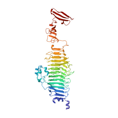
Crystal structure of phage P22 tailspike protein complexed with Salmonella sp. O-antigen receptors.

Steinbacher, S.Baxa, U.Miller, S.Weintraub, A.Seckler, R.Huber, R.

(1996) Proc Natl Acad Sci U S A 93: 10584-10588

  • DOI: https://doi.org/10.1073/pnas.93.20.10584
  • Primary Citation Related Structures: 
    1TYU, 1TYV, 1TYW, 1TYX

  • PubMed Abstract: 

    The O-antigenic repeating units of lipopolysaccharides from Salmonella serogroups A, B, and D1 serve as receptors for the phage P22 tailspike protein, which also has receptor destroying endoglycosidase (endorhamnosidase) activity, integrating the functions of both hemagglutinin and neuraminidase in influenza virus. Crystal structures of the tailspike protein in complex with oligosaccharides, comprising two O-antigenic repeating units from Salmonella typhimurium, Salmonella enteritidis, and Salmonella typhi 253Ty were determined at 1.8 A resolution. The active-site topology with Asp-392, Asp-395, and Glu-359 as catalytic residues was identified. Kinetics of binding and cleavage suggest a role of the receptor destroying endorhamnosidase activity primarily for detachment of newly assembled phages.


  • Organizational Affiliation
    • Abteilung Strukturforschung, Max-Planck-Institut für Biochemie, Martinsried, Germany.

Macromolecule Content 

  • Total Structure Weight: 60.84 kDa 
  • Atom Count: 4,407 
  • Modeled Residue Count: 543 
  • Deposited Residue Count: 554 
  • Unique protein chains: 1

Macromolecules

Find similar proteins by:|  3D Structure
Entity ID: 1
MoleculeChains  Sequence LengthOrganismDetailsImage
TAILSPIKE PROTEIN554Lederbergvirus P22Mutation(s): 0 
Gene Names: PHAGE P22 GENE 9
EC: 3.2.1
UniProt
Find proteins for P12528 (Salmonella phage P22)
Explore P12528 
Go to UniProtKB:  P12528
Entity Groups
Sequence Clusters30% Identity50% Identity70% Identity90% Identity95% Identity100% Identity
UniProt GroupP12528
Sequence Annotations
Expand
Reference Sequence

Oligosaccharides

Help  
Entity ID: 2
MoleculeChains Length2D Diagram GlycosylationD Interactions
alpha-D-galactopyranose-(1-2)-[alpha-D-Abequopyranose-(1-3)]alpha-D-mannopyranose-(1-4)-alpha-L-rhamnopyranose-(1-3)-alpha-D-galactopyranose-(1-2)-[alpha-D-Abequopyranose-(1-3)]alpha-D-mannopyranose-(1-4)-alpha-L-rhamnopyranose
B
8N/A
Glycosylation Resources
GlyTouCan: G56937DQ
GlyCosmos: G56937DQ

Experimental Data & Validation

Experimental Data

  • Method: X-RAY DIFFRACTION
  • Resolution: 1.80 Å
  • R-Value Work:  0.183 (Depositor) 
  • R-Value Observed: 0.183 (Depositor) 
Space Group: P 21 3
Unit Cell:
Length ( Å )Angle ( ˚ )
a = 120.9α = 90
b = 120.9β = 90
c = 120.9γ = 90
Software Package:
Software NamePurpose
X-PLORmodel building
X-PLORrefinement
MOSFLMdata reduction
X-PLORphasing

Structure Validation

View Full Validation Report


Entry History 

Deposition Data

Revision History  (Full details and data files)

  • Version 1.0: 1997-07-23
    Type: Initial release
  • Version 1.1: 2008-03-03
    Changes: Version format compliance
  • Version 1.2: 2011-07-13
    Changes: Derived calculations, Version format compliance
  • Version 2.0: 2020-07-29
    Type: Remediation
    Reason: Carbohydrate remediation
    Changes: Atomic model, Data collection, Derived calculations, Other, Structure summary
  • Version 2.1: 2024-02-14
    Changes: Data collection, Database references, Structure summary