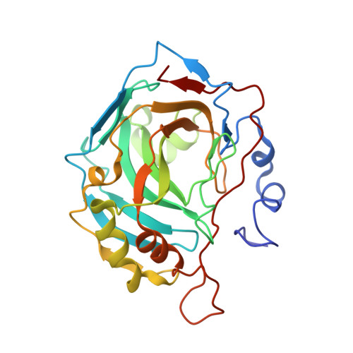
Crystal structure of human carbonic anhydrase II at 1.95 A resolution in complex with 667-coumate, a novel anti-cancer agent
Lloyd, M.D., Pederick, R.L., Natesh, R., Woo, L.W.L., Purohit, A., Reed, M.J., Acharya, K.R., Potter, B.V.L.(2005) Biochem J 385: 715-720
- PubMed: 15453828 Search on PubMedSearch on PubMed Central
- DOI: https://doi.org/10.1042/BJ20041037
- Primary Citation Related Structures:
1TTM - PubMed Abstract:
CA (carbonic anhydrase) catalyses the reversible hydration of carbon dioxide into bicarbonate, and at least 14 isoforms have been identified in vertebrates. The role of CA type II in maintaining the fluid and pH balance has made it an attractive drug target for the treatment of glaucoma and cancer. 667-coumate is a potent inhibitor of the novel oncology target steroid sulphatase and is currently in Phase 1 clinical trials for hormone-dependent breast cancer. It also inhibits CA II in vitro. In the present study, CA II was crystallized with 667-coumate and the structure was determined by X-ray crystallography at 1.95 A (1 A=0.1 nm) resolution. The structure reported here is the first for an inhibitor based on a coumarin ring and shows ligation of the sulphamate group to the active-site zinc at 2.15 A through a nitrogen anion. The first two rings of the coumarin moiety are bound within the hydrophobic binding site of CA II. Important residues contributing to binding include Val-121, Phe-131, Val-135, Leu-141, Leu-198 and Pro-202. The third seven-membered ring is more mobile and is located in the channel leading to the surface of the enzyme. Pharmacokinetic studies show enhanced stability of 667-coumate in vivo and this has been ascribed to binding of CA II in erythrocytes. This result provides a structural basis for the stabilization and long half-life of 667-coumate in blood compared with its rapid disappearance in plasma, and suggests that reversible binding of inhibitors to CA may be a general method of delivering this type of labile drug.
- Medicinal Chemistry, Department of Pharmacy and Pharmacology, University of Bath, Claverton Down, Bath BA2 7AY, UK. M.D.Lloyd@bath.ac.uk
Organizational Affiliation: