1TQ7

Crystal structure of the anticoagulant thrombin mutant W215A/E217A bound to PPACK


Experimental Data Snapshot

  • Method: X-RAY DIFFRACTION
  • Resolution: 2.40 Å
  • R-Value Free: 0.243 
  • R-Value Work: 0.205 
  • R-Value Observed: 0.205 

wwPDB Validation   3D Report Full Report


Ligand Structure Quality Assessment 


This is version 1.7 of the entry. See complete history


Literature

The anticoagulant thrombin mutant W215A/E217A has a collapsed primary specificity pocket

Pineda, A.O.Chen, Z.-W.Caccia, S.Cantwell, A.M.Savvides, S.N.Waksman, G.Mathews, F.S.Di Cera, E.

(2004) J Biol Chem 279: 39824-39828

  • DOI: https://doi.org/10.1074/jbc.M407272200
  • Primary Citation of Related Structures:  
    1TQ0, 1TQ7

  • PubMed Abstract: 

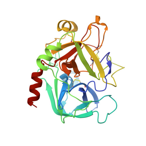
    The thrombin mutant W215A/E217A features a drastically impaired catalytic activity toward chromogenic and natural substrates but efficiently activates the anticoagulant protein C in the presence of thrombomodulin. As the remarkable anticoagulant properties of this mutant continue to be unraveled in preclinical studies, we solved the x-ray crystal structures of its free form and its complex with the active site inhibitor H-d-Phe-Pro-Arg-CH(2)Cl (PPACK). The PPACK-bound structure of W215A/E217A is identical to the structure of the PPACK-bound slow form of thrombin. On the other hand, the structure of the free form reveals a collapse of the 215-217 strand that crushes the primary specificity pocket. The collapse results from abrogation of the stacking interaction between Phe-227 and Trp-215 and the polar interactions of Glu-217 with Thr-172 and Lys-224. Other notable changes are a rotation of the carboxylate group of Asp-189, breakage of the H-bond between the catalytic residues Ser-195 and His-57, breakage of the ion pair between Asp-222 and Arg-187, and significant disorder in the 186- and 220-loops that define the Na(+) site. These findings explain the impaired catalytic activity of W215A/E217A and demonstrate that the analysis of the molecular basis of substrate recognition by thrombin and other proteases requires crystallization of both the free and bound forms of the enzyme.


  • Organizational Affiliation

    Department of Biochemistry and Molecular Biophysics, Washington University School of Medicine, St. Louis, Missouri 63110, USA.


Macromolecules
Find similar proteins by:  (by identity cutoff)  |  3D Structure
Entity ID: 1
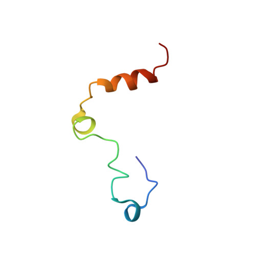
MoleculeChains Sequence LengthOrganismDetailsImage
Prothrombin44Homo sapiensMutation(s): 0 
Gene Names: F2
EC: 3.4.21.5
UniProt & NIH Common Fund Data Resources
Find proteins for P00734 (Homo sapiens)
Explore P00734 
Go to UniProtKB:  P00734
PHAROS:  P00734
GTEx:  ENSG00000180210 
Entity Groups  
Sequence Clusters30% Identity50% Identity70% Identity90% Identity95% Identity100% Identity
UniProt GroupP00734
Sequence Annotations
Expand
  • Reference Sequence
Find similar proteins by:  (by identity cutoff)  |  3D Structure
Entity ID: 2
MoleculeChains Sequence LengthOrganismDetailsImage
Prothrombin257Homo sapiensMutation(s): 2 
Gene Names: F2
EC: 3.4.21.5
UniProt & NIH Common Fund Data Resources
Find proteins for P00734 (Homo sapiens)
Explore P00734 
Go to UniProtKB:  P00734
PHAROS:  P00734
GTEx:  ENSG00000180210 
Entity Groups  
Sequence Clusters30% Identity50% Identity70% Identity90% Identity95% Identity100% Identity
UniProt GroupP00734
Sequence Annotations
Expand
  • Reference Sequence
Biologically Interesting Molecules (External Reference) 1 Unique
Experimental Data & Validation

Experimental Data

  • Method: X-RAY DIFFRACTION
  • Resolution: 2.40 Å
  • R-Value Free: 0.243 
  • R-Value Work: 0.205 
  • R-Value Observed: 0.205 
  • Space Group: P 21 21 21
Unit Cell:
Length ( Å )Angle ( ˚ )
a = 51.11α = 90
b = 73.83β = 90
c = 87.33γ = 90
Software Package:
Software NamePurpose
CNSrefinement
DENZOdata reduction
SCALEPACKdata scaling
CNSphasing

Structure Validation

View Full Validation Report


Ligand Structure Quality Assessment 


Entry History 

Deposition Data

Revision History  (Full details and data files)

  • Version 1.0: 2004-08-03
    Type: Initial release
  • Version 1.1: 2008-04-30
    Changes: Version format compliance
  • Version 1.2: 2011-07-13
    Changes: Atomic model, Database references, Derived calculations, Non-polymer description, Structure summary, Version format compliance
  • Version 1.3: 2013-02-27
    Changes: Other
  • Version 1.4: 2020-07-29
    Type: Remediation
    Reason: Carbohydrate remediation
    Changes: Data collection, Derived calculations, Structure summary
  • Version 1.5: 2021-10-27
    Changes: Database references, Structure summary
  • Version 1.6: 2023-08-23
    Changes: Data collection, Refinement description
  • Version 1.7: 2024-03-13
    Changes: Source and taxonomy, Structure summary