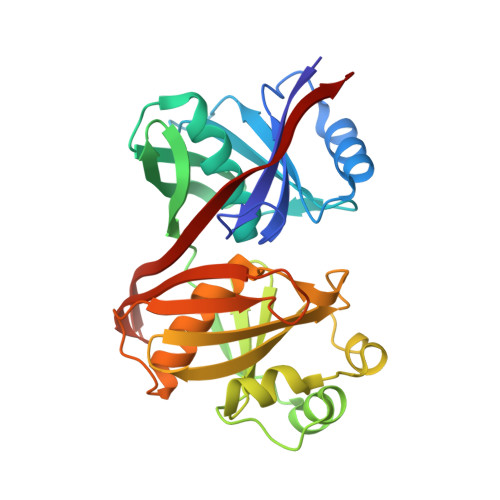
Structure and function of the phenazine biosynthesis protein PhzF from Pseudomonas fluorescens 2-79
Parsons, J.F., Song, F., Parsons, L., Calabrese, K., Eisenstein, E., Ladner, J.E.(2004) Biochemistry 43: 12427-12435
- PubMed: 15449932 Search on PubMed
- DOI: https://doi.org/10.1021/bi049059z
- Primary Citation Related Structures:
1T6K - PubMed Abstract:
Phenazines, including pyocyanin and iodonin, are biologically active compounds that are believed to confer producing organisms with a competitive growth advantage, and also are thought to be virulence factors in certain diseases including cystic fibrosis. The basic, tricyclic phenazine ring system is synthesized in a series of poorly characterized steps by enzymes encoded in a seven-gene cistron in Pseudomonas and other organisms. Despite the biological importance of these compounds, and our understanding of their mode of action, the biochemistry and mechanisms of phenazine biosynthesis are not well resolved. Here we report the 1.8 A crystal structure of PhzF, a key enzyme in phenazine biosynthesis, solved by molecular replacement. PhzF is structurally similar to the lysine biosynthetic enzyme diaminopimelate epimerase, sharing an unusual fold consisting of two nearly identical domains with the active site located in an occluded cleft between the domains. Unlike diaminopimelate epimerase, PhzF is a dimer in solution. The two apparently independent active sites open toward opposite sides of the dimer and are occupied by sulfate ions in the structure. In vitro experiments using a mixture of purified PhzF, -A, -B, and -G confirm that phenazine-1-carboxylic acid (PCA) is readily produced from trans-2,3-dihydro-3-hydroxyanthranilic acid (DHHA) without aid of other cellular factors. PhzA, -B, and -G have no activity toward DHHA. However, in the presence of PhzF, individually or in combinations, they accelerate the formation of PCA from DHHA and therefore appear to function after the action of PhzF. Surprisingly, PhzF is itself capable of producing PCA, albeit slowly, from DHHA. These observations suggest that PhzF catalyzes the initial step in the conversion of DHHA to PCA, probably via a rearrangement reaction yielding the more reactive 3-oxo analogue of DHHA, and that subsequent steps can occur spontaneously. A hypothetical model for how DHHA binds to the PhzF active site suggests that Glu45 and Asp208 could act as general acid-base catalysts in a rearrangement reaction. Given that four reactions lie between DHHA and PCA, ketone formation, ring formation, decarboxylation, and oxidation, we hypothesize that the similar PhzA and -B proteins catalyze ring formation and thus may be more than noncatalytic accessory proteins. PhzG is almost certainly an oxidase and is predicted to catalyze the final oxidation/aromatization reaction.
- Center for Advanced Research in Biotechnology, University of Maryland Biotechnology Institute, National Institute of Standards and Technology, 9600 Gudelsky Drive, Rockville, Maryland 20850, USA.
Organizational Affiliation: