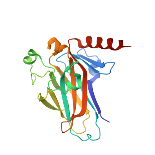
1T4W

Structural Differences in the DNA Binding Domains of Human p53 and its C. elegans Ortholog Cep-1: Structure of C. elegans Cep-1


Experimental Data Snapshot

  • Method: X-RAY DIFFRACTION
  • Resolution: 2.10 Å
  • R-Value Free: 0.234 
  • R-Value Work: 0.176 
  • R-Value Observed: 0.225 

wwPDB Validation   3D Report Full Report


This is version 1.3 of the entry. See complete history


Literature

Structural Differences in the DNA Binding Domains of Human p53 and Its C. elegans Ortholog Cep-1.

Huyen, Y.Jeffrey, P.D.Derry, W.B.Rothman, J.H.Pavletich, N.P.Stavridi, E.S.Halazonetis, T.D.

(2004) Structure 12: 1237-1243

  • DOI: https://doi.org/10.1016/j.str.2004.05.007
  • Primary Citation of Related Structures:  
    1T4W

  • PubMed Abstract: 

    The DNA binding domains of human p53 and Cep-1, its C. elegans ortholog, recognize essentially identical DNA sequences despite poor sequence similarity. We solved the three-dimensional structure of the Cep-1 DNA binding domain in the absence of DNA and compared it to that of human p53. The two domains have similar overall folds. However, three loops, involved in DNA and Zn binding in human p53, contain small alpha helices in Cep-1. The alpha helix in loop L3 of Cep-1 orients the side chains of two conserved arginines toward DNA; in human p53, both arginines are mutation hotspots, but only one contacts DNA. The alpha helix in loop L1 of Cep-1 repositions the entire loop, making it unlikely for residues of this loop to contact bases in the major groove of DNA, as occurs in human p53. Thus, during evolution there have been considerable changes in the structure of the p53 DNA binding domain.


  • Organizational Affiliation

    Wistar Institute, Philadelphia, PA 19104, USA.


Macromolecules
Find similar proteins by:  (by identity cutoff)  |  3D Structure
Entity ID: 1
MoleculeChains Sequence LengthOrganismDetailsImage
C.Elegans p53 tumor suppressor-like transcription factor196Caenorhabditis elegansMutation(s): 0 
UniProt
Find proteins for Q20646 (Caenorhabditis elegans)
Explore Q20646 
Go to UniProtKB:  Q20646
Entity Groups  
Sequence Clusters30% Identity50% Identity70% Identity90% Identity95% Identity100% Identity
UniProt GroupQ20646
Sequence Annotations
Expand
  • Reference Sequence
Small Molecules
Ligands 1 Unique
IDChains Name / Formula / InChI Key2D Diagram3D Interactions
ZN
Query on ZN

Download Ideal Coordinates CCD File 
B [auth A]ZINC ION
Zn
PTFCDOFLOPIGGS-UHFFFAOYSA-N
Experimental Data & Validation

Experimental Data

  • Method: X-RAY DIFFRACTION
  • Resolution: 2.10 Å
  • R-Value Free: 0.234 
  • R-Value Work: 0.176 
  • R-Value Observed: 0.225 
  • Space Group: P 21 21 21
Unit Cell:
Length ( Å )Angle ( ˚ )
a = 39.36α = 90
b = 68.43β = 90
c = 88.18γ = 90
Software Package:
Software NamePurpose
CNSrefinement
DENZOdata reduction
SCALEPACKdata scaling
MLPHAREphasing

Structure Validation

View Full Validation Report


Entry History 

Deposition Data

Revision History  (Full details and data files)

  • Version 1.0: 2004-07-20
    Type: Initial release
  • Version 1.1: 2008-04-30
    Changes: Version format compliance
  • Version 1.2: 2011-07-13
    Changes: Version format compliance
  • Version 1.3: 2024-02-14
    Changes: Data collection, Database references, Derived calculations