Structural Differences in the DNA Binding Domains of Human p53 and Its C. elegans Ortholog Cep-1.
Huyen, Y., Jeffrey, P.D., Derry, W.B., Rothman, J.H., Pavletich, N.P., Stavridi, E.S., Halazonetis, T.D.(2004) Structure 12: 1237-1243
- PubMed: 15242600 Search on PubMed
- DOI: https://doi.org/10.1016/j.str.2004.05.007
- Primary Citation Related Structures:
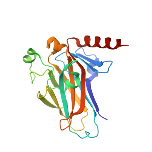
1T4W - PubMed Abstract:
The DNA binding domains of human p53 and Cep-1, its C. elegans ortholog, recognize essentially identical DNA sequences despite poor sequence similarity. We solved the three-dimensional structure of the Cep-1 DNA binding domain in the absence of DNA and compared it to that of human p53. The two domains have similar overall folds. However, three loops, involved in DNA and Zn binding in human p53, contain small alpha helices in Cep-1. The alpha helix in loop L3 of Cep-1 orients the side chains of two conserved arginines toward DNA; in human p53, both arginines are mutation hotspots, but only one contacts DNA. The alpha helix in loop L1 of Cep-1 repositions the entire loop, making it unlikely for residues of this loop to contact bases in the major groove of DNA, as occurs in human p53. Thus, during evolution there have been considerable changes in the structure of the p53 DNA binding domain.
- Wistar Institute, Philadelphia, PA 19104, USA.
Organizational Affiliation: