1T3T

Structure of Formylglycinamide synthetase


Experimental Data Snapshot

  • Method: X-RAY DIFFRACTION
  • Resolution: 1.90 Å
  • R-Value Free: 0.206 
  • R-Value Work: 0.188 
  • R-Value Observed: 0.188 

wwPDB Validation   3D Report Full Report


Ligand Structure Quality Assessment 


This is version 1.2 of the entry. See complete history


Literature

Domain Organization of Salmonella typhimurium Formylglycinamide Ribonucleotide Amidotransferase Revealed by X-ray crystallography

Anand, R.Hoskin, A.A.Stubbe, J.Ealick, S.E.

(2004) Biochemistry 43: 10328-10342

  • DOI: https://doi.org/10.1021/bi0491301
  • Primary Citation of Related Structures:  
    1T3T

  • PubMed Abstract: 

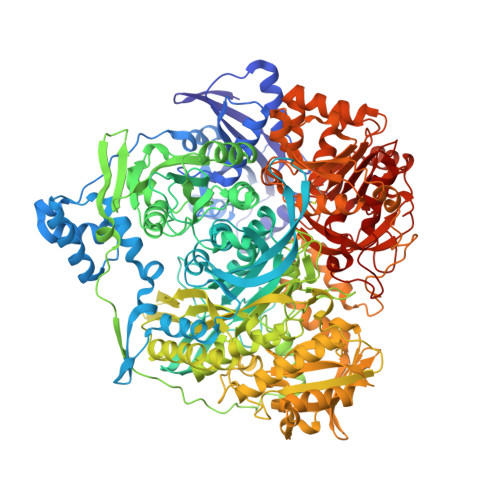
    Formylglycinamide ribonucleotide amidotransferase (FGAR-AT) catalyzes the ATP-dependent conversion of formylglycinamide ribonucleotide (FGAR) and glutamine to formylglycinamidine ribonucleotide (FGAM), ADP, P(i), and glutamate in the fourth step of the purine biosynthetic pathway. In eukaryotes and Gram-negative bacteria, FGAR-AT is encoded by the purL gene as a multidomain protein with a molecular mass of about 140 kDa. In Gram-positive bacteria and archaebacteria FGAR-AT is a complex of three proteins: PurS, PurL, and PurQ. We have determined the structure of FGAR-AT (PurL) from Salmonella typhimurium at 1.9 A resolution using X-ray crystallography. PurL is the last remaining enzyme in the purine biosynthetic pathway to have its structure determined. The structure reveals four domains: an N-terminal domain structurally homologous to a PurS dimer, a linker region, an FGAM synthetase domain homologous to an aminoimidazole ribonucleotide synthetase (PurM) dimer, and a triad glutaminase domain. The domains are intricately linked by interdomain interactions and peptide connectors. The fold common to PurM and the central region of PurL represents a superfamily for which HypE, SelD, and ThiL are predicted to be members. A structural ADP molecule was found bound to a site related to the putative active site by pseudo-2-fold symmetry and two sulfate ions were found at the putative active site. These observations and the structural similarities between PurM and StPurL were used to model the substrates FGAR and ATP in the StPurL active site. A glutamylthioester intermediate was found in the glutaminase domain at Cys1135. The N-terminal (PurS-like) domain is hypothesized to form the putative channel through which ammonia passes from the glutaminase domain to the FGAM synthetase domain.


  • Organizational Affiliation

    Department of Chemistry and Chemical Biology, Cornell University, Ithaca, New York 14853, USA.


Macromolecules
Find similar proteins by:  (by identity cutoff)  |  3D Structure
Entity ID: 1
MoleculeChains Sequence LengthOrganismDetailsImage
Phosphoribosylformylglycinamidine synthase1,303Salmonella enterica subsp. enterica serovar TyphimuriumMutation(s): 1 
Gene Names: PURLSTM2565
EC: 6.3.5.3
UniProt
Find proteins for P74881 (Salmonella typhimurium (strain LT2 / SGSC1412 / ATCC 700720))
Explore P74881 
Go to UniProtKB:  P74881
Entity Groups  
Sequence Clusters30% Identity50% Identity70% Identity90% Identity95% Identity100% Identity
UniProt GroupP74881
Sequence Annotations
Expand
  • Reference Sequence
Small Molecules
Ligands 3 Unique
IDChains Name / Formula / InChI Key2D Diagram3D Interactions
ADP
Query on ADP

Download Ideal Coordinates CCD File 
K [auth A]ADENOSINE-5'-DIPHOSPHATE
C10 H15 N5 O10 P2
XTWYTFMLZFPYCI-KQYNXXCUSA-N
SO4
Query on SO4

Download Ideal Coordinates CCD File 
E [auth A]
F [auth A]
G [auth A]
H [auth A]
I [auth A]
E [auth A],
F [auth A],
G [auth A],
H [auth A],
I [auth A],
J [auth A]
SULFATE ION
O4 S
QAOWNCQODCNURD-UHFFFAOYSA-L
MG
Query on MG

Download Ideal Coordinates CCD File 
B [auth A],
C [auth A],
D [auth A]
MAGNESIUM ION
Mg
JLVVSXFLKOJNIY-UHFFFAOYSA-N
Modified Residues  1 Unique
IDChains TypeFormula2D DiagramParent
CYG
Query on CYG
A
L-PEPTIDE LINKINGC8 H14 N2 O5 SCYS
Binding Affinity Annotations 
IDSourceBinding Affinity
ADP PDBBind:  1T3T Ki: 1.10e+4 (nM) from 1 assay(s)
Experimental Data & Validation

Experimental Data

  • Method: X-RAY DIFFRACTION
  • Resolution: 1.90 Å
  • R-Value Free: 0.206 
  • R-Value Work: 0.188 
  • R-Value Observed: 0.188 
  • Space Group: P 65
Unit Cell:
Length ( Å )Angle ( ˚ )
a = 145.806α = 90
b = 145.806β = 90
c = 141.353γ = 120
Software Package:
Software NamePurpose
CNSrefinement
MOSFLMdata reduction
CCP4data scaling
CNSphasing

Structure Validation

View Full Validation Report


Ligand Structure Quality Assessment 


Entry History 

Deposition Data

Revision History  (Full details and data files)

  • Version 1.0: 2004-09-14
    Type: Initial release
  • Version 1.1: 2008-04-30
    Changes: Version format compliance
  • Version 1.2: 2011-07-13
    Changes: Version format compliance