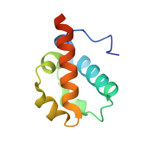
1SV4

Crystal Structure of Yan-SAM


Experimental Data Snapshot

  • Method: X-RAY DIFFRACTION
  • Resolution: 2.15 Å
  • R-Value Free: 0.249 
  • R-Value Work: 0.218 
  • R-Value Observed: 0.218 

Starting Model: experimental
View more details

wwPDB Validation   3D Report Full Report


This is version 1.4 of the entry. See complete history


Literature

Derepression by depolymerization; structural insights into the regulation of yan by mae.

Qiao, F.Song, H.Kim, C.A.Sawaya, M.R.Hunter, J.B.Gingery, M.Rebay, I.Courey, A.J.Bowie, J.U.

(2004) Cell 118: 163-173

  • DOI: https://doi.org/10.1016/j.cell.2004.07.010
  • Primary Citation of Related Structures:  
    1SV0, 1SV4

  • PubMed Abstract: 

    Yan, an ETS family transcriptional repressor, is regulated by receptor tyrosine kinase signaling via the Ras/MAPK pathway. Phosphorylation and downregulation of Yan is facilitated by a protein called Mae. Yan and Mae interact through their SAM domains. We find that repression by Yan requires the formation of a higher order structure mediated by Yan-SAM polymerization. Moreover, a crystal structure of the Yan-SAM/Mae-SAM complex shows that Mae-SAM specifically recognizes a surface on Yan-SAM that is also required for Yan-SAM polymerization. Mae-SAM binds to Yan-SAM with approximately 1000-fold higher affinity than Yan-SAM binds to itself and can effectively depolymerize Yan-SAM. Mutations on Mae that specifically disrupt its SAM domain-dependent interactions with Yan disable the derepression function of Mae in vivo. Depolymerization of Yan by Mae represents a novel mechanism of transcriptional control that sensitizes Yan for regulation by receptor tyrosine kinases.


  • Organizational Affiliation

    UCLA-DOE Institute of Genomics and Proteomics, Molecular Biology Institute, Department of Chemistry and Biochemistry, University of California, Los Angeles 90095, USA.


Macromolecules
Find similar proteins by:  (by identity cutoff)  |  3D Structure
Entity ID: 1
MoleculeChains Sequence LengthOrganismDetailsImage
Ets DNA-binding protein pokkuri
A, B
85Drosophila melanogasterMutation(s): 1 
Gene Names: AOPPOKYANCG3166
UniProt
Find proteins for Q01842 (Drosophila melanogaster)
Explore Q01842 
Go to UniProtKB:  Q01842
Entity Groups  
Sequence Clusters30% Identity50% Identity70% Identity90% Identity95% Identity100% Identity
UniProt GroupQ01842
Sequence Annotations
Expand
  • Reference Sequence
Experimental Data & Validation

Experimental Data

  • Method: X-RAY DIFFRACTION
  • Resolution: 2.15 Å
  • R-Value Free: 0.249 
  • R-Value Work: 0.218 
  • R-Value Observed: 0.218 
  • Space Group: C 1 2 1
Unit Cell:
Length ( Å )Angle ( ˚ )
a = 76.51α = 90
b = 45.41β = 108.47
c = 55.6γ = 90
Software Package:
Software NamePurpose
CNSrefinement
DENZOdata reduction
SCALEPACKdata scaling
EPMRphasing

Structure Validation

View Full Validation Report


Entry History 

Deposition Data

Revision History  (Full details and data files)

  • Version 1.0: 2004-07-27
    Type: Initial release
  • Version 1.1: 2008-04-30
    Changes: Version format compliance
  • Version 1.2: 2011-07-13
    Changes: Version format compliance
  • Version 1.3: 2021-10-27
    Changes: Database references
  • Version 1.4: 2023-08-23
    Changes: Data collection, Refinement description