1SKN | pdb_00001skn

THE BINDING DOMAIN OF SKN-1 IN COMPLEX WITH DNA: A NEW DNA-BINDING MOTIF


Experimental Data Snapshot

  • Method: X-RAY DIFFRACTION
  • Resolution: 2.50 Å
  • R-Value Free: 
    0.337 (Depositor), 0.325 (DCC) 
  • R-Value Work: 
    0.220 (Depositor), 0.227 (DCC) 
  • R-Value Observed: 
    0.240 (Depositor) 

wwPDB Validation 3D Report Full Report

Validation slider image for 1SKN

Ligand Structure Quality Assessment 


This is version 1.4 of the entry. See complete history

Literature

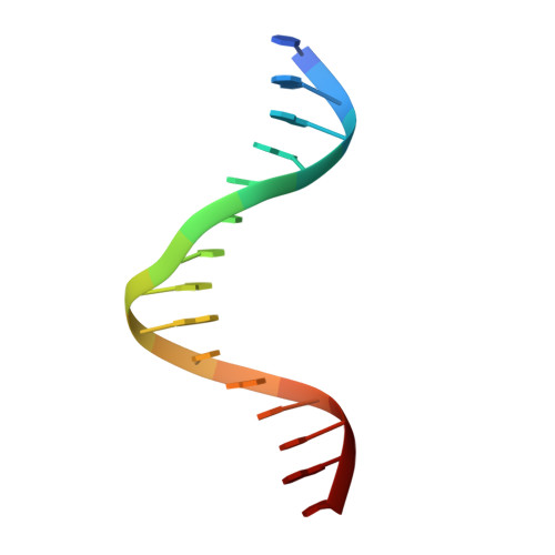
A new DNA-binding motif in the Skn-1 binding domain-DNA complex.

Rupert, P.B.Daughdrill, G.W.Bowerman, B.Matthews, B.W.

(1998) Nat Struct Biol 5: 484-491

  • DOI: https://doi.org/10.1038/nsb0698-484
  • Primary Citation Related Structures: 
    1SKN

  • PubMed Abstract: 

    The DNA-binding domain of Skn-1, a developmental transcription factor that specifies mesoderm in C. elegans, is shown by X-ray crystallography to have a novel fold in which a compact, monomeric, four-helix unit organizes two DNA-contact elements. At the C-terminus, a helix extends from the domain to occupy the major groove of DNA in a manner similar to bZip proteins. Skn-1, however, lacks the leucine zipper found in all bZips. Additional contacts with the DNA are made by a short basic segment at the N-terminus of the domain, reminiscent of the 'homeodomain arm'.


  • Organizational Affiliation
    • Institute of Molecular Biology, Department of Chemistry, University of Oregon, Eugene 97403, USA.

Macromolecule Content 

  • Total Structure Weight: 20.4 kDa 
  • Atom Count: 1,263 
  • Modeled Residue Count: 104 
  • Deposited Residue Count: 122 
  • Unique protein chains: 1
  • Unique nucleic acid chains: 2

Macromolecules


Find similar proteins by:|  3D Structure
Entity ID: 3
MoleculeChains  Sequence LengthOrganismDetailsImage
DNA-BINDING DOMAIN OF SKN-1C [auth P]92Caenorhabditis elegansMutation(s): 0 
Gene Names: SKN-1
UniProt
Find proteins for P34707 (Caenorhabditis elegans)
Explore P34707 
Go to UniProtKB:  P34707
Entity Groups
Sequence Clusters30% Identity50% Identity70% Identity90% Identity95% Identity100% Identity
UniProt GroupP34707
Sequence Annotations
Expand
Reference Sequence
Find similar nucleic acids by:  Sequence
Entity ID: 1
MoleculeChains LengthOrganismImage
DNA (5'-D(*TP*GP*AP*CP*AP*AP*TP*GP*TP*CP*AP*TP*CP*CP*C)-3')15N/A
Sequence Annotations
Expand
Reference Sequence
Find similar nucleic acids by:  Sequence
Entity ID: 2
MoleculeChains LengthOrganismImage
DNA (5'-D(*CP*AP*GP*GP*GP*AP*TP*GP*AP*CP*AP*TP*TP*GP*T)-3')15N/A
Sequence Annotations
Expand
Reference Sequence

Small Molecules

Ligands 1 Unique
IDChains Name / Formula / InChI Key2D Diagram3D Interactions
LDA

Query on LDA


Download:Ideal Coordinates CCD File
D [auth P]LAURYL DIMETHYLAMINE-N-OXIDE
C14 H31 N O
SYELZBGXAIXKHU-UHFFFAOYSA-N

Experimental Data & Validation

Experimental Data

  • Method: X-RAY DIFFRACTION
  • Resolution: 2.50 Å
  • R-Value Free:  0.337 (Depositor), 0.325 (DCC) 
  • R-Value Work:  0.220 (Depositor), 0.227 (DCC) 
  • R-Value Observed: 0.240 (Depositor) 
Space Group: C 2 2 21
Unit Cell:
Length ( Å )Angle ( ˚ )
a = 82.13α = 90
b = 93.39β = 90
c = 53.45γ = 90
Software Package:
Software NamePurpose
TNTrefinement
XTALVIEWrefinement
DENZOdata reduction
SCALEPACKdata scaling

Structure Validation

View Full Validation Report


Ligand Structure Quality Assessment 


Entry History 

Deposition Data

Revision History  (Full details and data files)

  • Version 1.0: 1998-06-24
    Type: Initial release
  • Version 1.1: 2008-05-22
    Changes: Version format compliance
  • Version 1.2: 2011-07-13
    Changes: Version format compliance
  • Version 1.3: 2019-07-17
    Changes: Data collection, Derived calculations, Refinement description
  • Version 1.4: 2024-02-14
    Changes: Data collection, Database references, Derived calculations