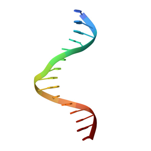
1SKN | pdb_00001skn

THE BINDING DOMAIN OF SKN-1 IN COMPLEX WITH DNA: A NEW DNA-BINDING MOTIF


Experimental Data Snapshot

  • Method: X-RAY DIFFRACTION
  • Resolution: 2.50 Å
  • R-Value Free: 
    0.337 (Depositor), 0.330 (DCC) 
  • R-Value Work: 
    0.220 (Depositor), 0.230 (DCC) 
  • R-Value Observed: 
    0.240 (Depositor) 

wwPDB Validation   3D Report Full Report


Ligand Structure Quality Assessment 

Created with Raphaël 2.3.0Worse 01 BetterLigand structure goodness of fit to experimental dataBest fitted LDAClick on this verticalbar to view details

This is version 1.4 of the entry. See complete history


Literature

A new DNA-binding motif in the Skn-1 binding domain-DNA complex.

Rupert, P.B.Daughdrill, G.W.Bowerman, B.Matthews, B.W.

(1998) Nat Struct Biol 5: 484-491

  • DOI: https://doi.org/10.1038/nsb0698-484
  • Primary Citation of Related Structures:  
    1SKN

  • PubMed Abstract: 

    The DNA-binding domain of Skn-1, a developmental transcription factor that specifies mesoderm in C. elegans, is shown by X-ray crystallography to have a novel fold in which a compact, monomeric, four-helix unit organizes two DNA-contact elements. At the C-terminus, a helix extends from the domain to occupy the major groove of DNA in a manner similar to bZip proteins. Skn-1, however, lacks the leucine zipper found in all bZips. Additional contacts with the DNA are made by a short basic segment at the N-terminus of the domain, reminiscent of the 'homeodomain arm'.


  • Organizational Affiliation

    Institute of Molecular Biology, Department of Chemistry, University of Oregon, Eugene 97403, USA.


Macromolecules

Find similar proteins by:  (by identity cutoff)  |  3D Structure
Entity ID: 3
MoleculeChains Sequence LengthOrganismDetailsImage
DNA-BINDING DOMAIN OF SKN-1C [auth P]92Caenorhabditis elegansMutation(s): 0 
Gene Names: SKN-1
UniProt
Find proteins for P34707 (Caenorhabditis elegans)
Explore P34707 
Go to UniProtKB:  P34707
Entity Groups  
Sequence Clusters30% Identity50% Identity70% Identity90% Identity95% Identity100% Identity
UniProt GroupP34707
Sequence Annotations
Expand
  • Reference Sequence
Small Molecules
Ligands 1 Unique
IDChains Name / Formula / InChI Key2D Diagram3D Interactions
LDA
Query on LDA

Download Ideal Coordinates CCD File 
D [auth P]LAURYL DIMETHYLAMINE-N-OXIDE
C14 H31 N O
SYELZBGXAIXKHU-UHFFFAOYSA-N
Experimental Data & Validation

Experimental Data

  • Method: X-RAY DIFFRACTION
  • Resolution: 2.50 Å
  • R-Value Free:  0.337 (Depositor), 0.330 (DCC) 
  • R-Value Work:  0.220 (Depositor), 0.230 (DCC) 
  • R-Value Observed: 0.240 (Depositor) 
Space Group: C 2 2 21
Unit Cell:
Length ( Å )Angle ( ˚ )
a = 82.13α = 90
b = 93.39β = 90
c = 53.45γ = 90
Software Package:
Software NamePurpose
TNTrefinement
XTALVIEWrefinement
DENZOdata reduction
SCALEPACKdata scaling

Structure Validation

View Full Validation Report


Ligand Structure Quality Assessment 

Created with Raphaël 2.3.0Worse 01 BetterLigand structure goodness of fit to experimental dataBest fitted LDAClick on this verticalbar to view details

Entry History 

Deposition Data

Revision History  (Full details and data files)

  • Version 1.0: 1998-06-24
    Type: Initial release
  • Version 1.1: 2008-05-22
    Changes: Version format compliance
  • Version 1.2: 2011-07-13
    Changes: Version format compliance
  • Version 1.3: 2019-07-17
    Changes: Data collection, Derived calculations, Refinement description
  • Version 1.4: 2024-02-14
    Changes: Data collection, Database references, Derived calculations