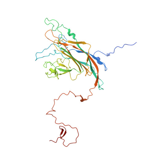
Crystal structures of murine polyomavirus in complex with straight-chain and branched-chain sialyloligosaccharide receptor fragments.
Stehle, T., Harrison, S.C.(1996) Structure 4: 183-194
- PubMed: 8805524 Search on PubMed
- DOI: https://doi.org/10.1016/s0969-2126(96)00021-4
- Primary Citation Related Structures:
1SID, 1SIE - PubMed Abstract:
Murine polyomavirus recognizes (alpha2,3)-linked alpha-5-N-acetylneuraminic acid (sialic acid) on the surface of susceptible cells. While all strains bind to straight-chain receptors terminating in (alpha2,3)-linked sialic acid, some strains also bind to branched oligosaccharides that carry a second, (alpha2,6)-linked sialic acid. The ability to bind to these branched-chain receptors correlates with a single amino acid mutation at position 91 on the outer surface of the major capsid protein, VP1, and with a significant decrease in tumorigenicity. We have determined the structures of polyomavirus strain P16, which bears a glycine at position 91, in complex with model compounds for both straight-chain and branched-chain sialoglycoconjugates. The structures have been refined to a resolution of 3.65 degree. The ligands bind to a shallow groove on the surface of VP1. The sialic acid-(alpha2,3)-galactose moiety, which is common to both compounds, has specific and identical contacts. The additional (alpha2,6)-linked sialic acid moiety of the branched-chain receptor fragment fits into a surface pocket, but it has high thermal factors and does not form hydrogen bonds to groups on VP1. Data collected from crystals soaked at different oligosaccharide concentrations establish that both receptor fragments have similar, low affinities (dissociation constants in the range 5-10 mM) for the P16 virus, consistent with the interactions seen in the two complexes. The oligosaccharide-binding groove is complementary to the shape of the bound glycan, but there are relatively few hydrogen bonds between glycan and protein. Thus, the nature of the glycosidic linkages appears to be the principal determinant of specificity, rather than the position of particular hydroxyl groups. The low receptor affinity may be important for avoiding inhibition of viral release by retention on surface receptors of infected cells. Evidence suggests that strains with still greater pathogenicity are likely to have even weaker affinity.
- Howard Hughes Medical Institute and Department of Molecular and Cellular Biology, Harvard University, Cambridge, MA 02138, USA.
Organizational Affiliation: