1SGH

Moesin FERM domain bound to EBP50 C-terminal peptide


Experimental Data Snapshot

  • Method: X-RAY DIFFRACTION
  • Resolution: 3.50 Å
  • R-Value Free: 0.401 
  • R-Value Work: 0.338 

wwPDB Validation   3D Report Full Report


This is version 1.3 of the entry. See complete history


Literature

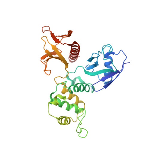
The EBP50-moesin interaction involves a binding site regulated by direct masking on the FERM domain

Finnerty, C.M.Chambers, D.Ingraffea, J.Faber, H.R.Karplus, P.A.Bretscher, A.

(2004) J Cell Sci 117: 1547-1552

  • DOI: https://doi.org/10.1242/jcs.01038
  • Primary Citation of Related Structures:  
    1SGH

  • PubMed Abstract: 
  • Members of the ezrin-radixin-moesin (ERM) protein family serve as regulated microfilament-membrane crosslinking proteins that, upon activation, bind the scaffolding protein ERM-phosphoprotein of 50 kDa (EBP50). Here we report a 3.5 A resolution diffraction analysis of a complex between the active moesin N-terminal FERM domain and a 38 residue peptide from the C terminus of EBP50 ...

    Members of the ezrin-radixin-moesin (ERM) protein family serve as regulated microfilament-membrane crosslinking proteins that, upon activation, bind the scaffolding protein ERM-phosphoprotein of 50 kDa (EBP50). Here we report a 3.5 A resolution diffraction analysis of a complex between the active moesin N-terminal FERM domain and a 38 residue peptide from the C terminus of EBP50. This crystallographic result, combined with sequence and structural comparisons, suggests that the C-terminal 11 residues of EBP50 binds as an alpha-helix at the same site occupied in the dormant monomer by the last 11 residues of the inhibitory moesin C-terminal tail. Biochemical support for this interpretation derives from in vitro studies showing that appropriate mutations in both the EBP50 tail peptide and the FERM domain reduce binding, and that a peptide representing just the C-terminal 14 residues of EBP50 also binds to moesin. Combined with the recent identification of the I-CAM-2 binding site on the ERM FERM domain (Hamada, K., Shimizu, T., Yonemura, S., Tsukita, S., and Hakoshima, T. (2003) EMBO J. 22, 502-514), this study reveals that the FERM domain contains two distinct binding sites for membrane-associated proteins. The contribution of each ligand to ERM function can now be dissected by making structure-based mutations that specifically affect the binding of each ligand.


    Organizational Affiliation

    Department of Molecular Biology and Genetics, Biotechnology Building, Cornell University, Ithaca, NY 14853, USA.


Macromolecules
Find similar proteins by:  (by identity cutoff)  |  3D Structure
Entity ID: 1
MoleculeChainsSequence LengthOrganismDetailsImage
Moesin297Homo sapiensMutation(s): 0 
Gene Names: MSN
UniProt & NIH Common Fund Data Resources
Find proteins for P26038 (Homo sapiens)
Explore P26038 
Go to UniProtKB:  P26038
PHAROS:  P26038
Entity Groups  
Sequence Clusters30% Identity50% Identity70% Identity90% Identity95% Identity100% Identity
UniProt GroupP26038
Protein Feature View
Expand
  • Reference Sequence
Find similar proteins by:  (by identity cutoff)  |  3D Structure
Entity ID: 2
MoleculeChainsSequence LengthOrganismDetailsImage
Ezrin-radixin-moesin binding phosphoprotein 5039N/AMutation(s): 0 
UniProt & NIH Common Fund Data Resources
Find proteins for O14745 (Homo sapiens)
Explore O14745 
Go to UniProtKB:  O14745
PHAROS:  O14745
Entity Groups  
Sequence Clusters30% Identity50% Identity70% Identity90% Identity95% Identity100% Identity
UniProt GroupO14745
Protein Feature View
Expand
  • Reference Sequence
Experimental Data & Validation

Experimental Data

  • Method: X-RAY DIFFRACTION
  • Resolution: 3.50 Å
  • R-Value Free: 0.401 
  • R-Value Work: 0.338 
  • Space Group: C 1 2 1
Unit Cell:
Length ( Å )Angle ( ˚ )
a = 127.6α = 90
b = 70.5β = 106.1
c = 62.8γ = 90
Software Package:
Software NamePurpose
MOSFLMdata reduction
SCALAdata scaling
CNSrefinement
CCP4data scaling
CNSphasing

Structure Validation

View Full Validation Report


Entry History 

Deposition Data

Revision History  (Full details and data files)

  • Version 1.0: 2004-06-29
    Type: Initial release
  • Version 1.1: 2008-04-29
    Changes: Version format compliance
  • Version 1.2: 2011-07-13
    Changes: Version format compliance
  • Version 1.3: 2023-08-23
    Changes: Data collection, Database references, Refinement description