1S5P | pdb_00001s5p

Structure and substrate binding properties of cobB, a Sir2 homolog protein deacetylase from Eschericia coli.


Experimental Data Snapshot

  • Method: X-RAY DIFFRACTION
  • Resolution: 1.96 Å
  • R-Value Free: 
    0.270 (Depositor), 0.244 (DCC) 
  • R-Value Work: 
    0.234 (Depositor), 0.231 (DCC) 
  • R-Value Observed: 
    0.234 (Depositor) 

wwPDB Validation 3D Report Full Report

Validation slider image for 1S5P

This is version 1.3 of the entry. See complete history

Literature

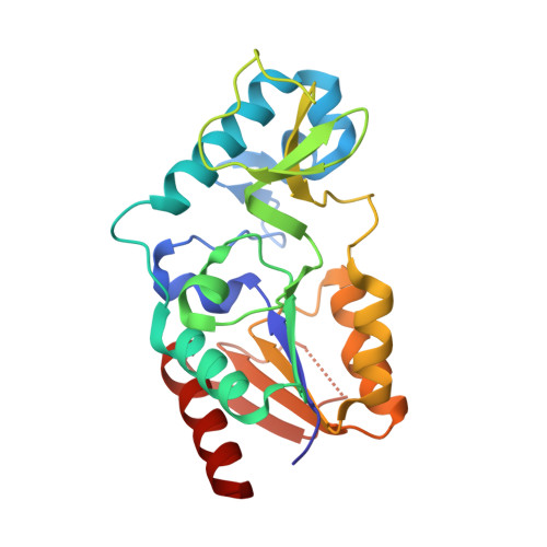
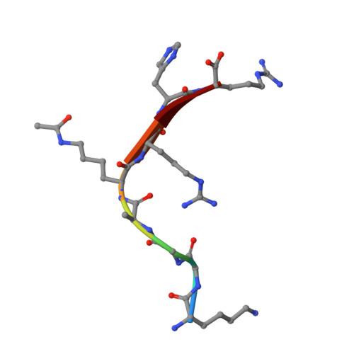
Structure and Substrate Binding Properties of cobB, a Sir2 Homolog Protein Deacetylase from Eschericia coli.

Zhao, K.Chai, X.Marmorstein, R.

(2004) J Mol Biology 337: 731-741

  • DOI: https://doi.org/10.1016/j.jmb.2004.01.060
  • Primary Citation Related Structures: 
    1S5P

  • PubMed Abstract: 

    Sirtuins are NAD+-dependent protein deacetylase enzymes that are broadly conserved from bacteria to human, and have been implicated to play important roles in gene regulation, metabolism and longevity. cobB is a bacterial sirtuin that deacetylates acetyl-CoA synthetase (Acs) at an active site lysine to stimulate its enzymatic activity. Here, we report the structure of cobB bound to an acetyl-lysine containing non-cognate histone H4 substrate. A comparison with the previously reported archaeal and eukaryotic sirtuin structures reveals the greatest variability in a small zinc-binding domain implicated to play a particularly important role in substrate-specific binding by the sirtuin proteins. Comparison of the cobB/histone H4 complex with other sirtuin proteins in complex with acetyl-lysine containing substrates, further suggests that contacts to the acetyl-lysine side-chain and beta-sheet interactions with residues directly C-terminal to the acetyl-lysine represent conserved features of sirtuin-substrate recognition. Isothermal titration calorimetry studies were used to compare the affinity of cobB for a variety of cognate and non-cognate acetyl-lysine-bearing peptides revealing an exothermic reaction with relatively little discrimination between substrates. In contrast, similar studies employing intact acetylated Acs protein as a substrate reveal a binding reaction that is endothermic, suggesting that cobB recognition of substrate involves a burial of hydrophobic surface and/or structural rearrangement involving substrate regions distal to the acetyl-lysine-binding site. Together, these studies suggest that substrate-specific binding by sirtuin proteins involves contributions from the zinc-binding domain of the enzyme and substrate regions distal to the acetyl-lysine-binding site.


  • Organizational Affiliation
    • The Wistar Institute, University of Pennsylvania, Philadelphia, PA 19104, USA.

Macromolecule Content 

  • Total Structure Weight: 27.11 kDa 
  • Atom Count: 2,044 
  • Modeled Residue Count: 233 
  • Deposited Residue Count: 243 
  • Unique protein chains: 2

Macromolecules

Find similar proteins by:|  3D Structure
Entity ID: 1
MoleculeChains  Sequence LengthOrganismDetailsImage
NAD-dependent deacetylase235Escherichia coliMutation(s): 0 
Gene Names: NPDACOBBB1120
EC: 3.5.1 (PDB Primary Data), 2.3.1.286 (UniProt)
UniProt
Find proteins for P75960 (Escherichia coli (strain K12))
Explore P75960 
Go to UniProtKB:  P75960
Entity Groups
Sequence Clusters30% Identity50% Identity70% Identity90% Identity95% Identity100% Identity
UniProt GroupP75960
Sequence Annotations
Expand
Reference Sequence
Find similar proteins by:  Sequence   |   3D Structure  
Entity ID: 2
MoleculeChains  Sequence LengthOrganismDetailsImage
HISTONE H4 (RESIDUES 12-19)8N/AMutation(s): 0 

Small Molecules

Ligands 1 Unique
IDChains Name / Formula / InChI Key2D Diagram3D Interactions
ZN

Query on ZN


Download:Ideal Coordinates CCD File
C [auth A]ZINC ION
Zn
PTFCDOFLOPIGGS-UHFFFAOYSA-N
Modified Residues  1 Unique
IDChains TypeFormula2D DiagramParent
ALY
Query on ALY
B
L-PEPTIDE LINKINGC8 H16 N2 O3LYS

Experimental Data & Validation

Experimental Data

  • Method: X-RAY DIFFRACTION
  • Resolution: 1.96 Å
  • R-Value Free:  0.270 (Depositor), 0.244 (DCC) 
  • R-Value Work:  0.234 (Depositor), 0.231 (DCC) 
  • R-Value Observed: 0.234 (Depositor) 
Space Group: P 41 21 2
Unit Cell:
Length ( Å )Angle ( ˚ )
a = 93.911α = 90
b = 93.911β = 90
c = 61.488γ = 90
Software Package:
Software NamePurpose
CNSrefinement
HKL-2000data reduction
SCALEPACKdata scaling
CNSphasing

Structure Validation

View Full Validation Report


Entry History 

Deposition Data

Revision History  (Full details and data files)

  • Version 1.0: 2004-03-23
    Type: Initial release
  • Version 1.1: 2008-04-29
    Changes: Version format compliance
  • Version 1.2: 2011-07-13
    Changes: Version format compliance
  • Version 1.3: 2024-10-09
    Changes: Data collection, Database references, Derived calculations, Structure summary