1RTJ | pdb_00001rtj

MECHANISM OF INHIBITION OF HIV-1 REVERSE TRANSCRIPTASE BY NON-NUCLEOSIDE INHIBITORS


Experimental Data Snapshot

  • Method: X-RAY DIFFRACTION
  • Resolution: 2.35 Å
  • R-Value Work: 
    0.219 (Depositor) 
  • R-Value Observed: 
    0.219 (Depositor) 

wwPDB Validation 3D Report Full Report

Validation slider image for 1RTJ

This is version 1.4 of the entry. See complete history

Literature

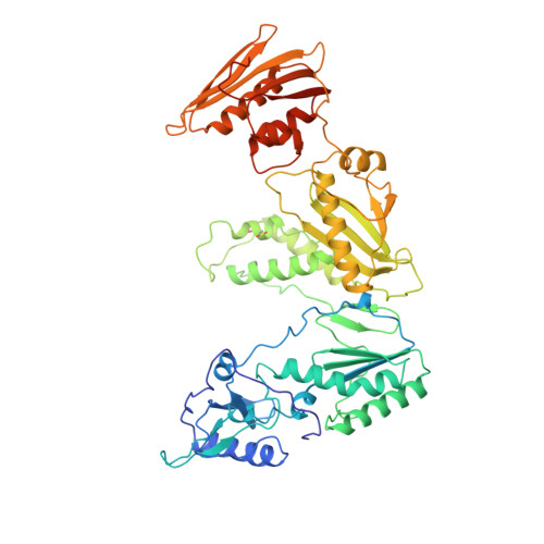
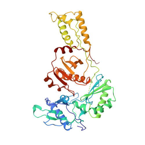
Mechanism of inhibition of HIV-1 reverse transcriptase by non-nucleoside inhibitors.

Esnouf, R.Ren, J.Ross, C.Jones, Y.Stammers, D.Stuart, D.

(1995) Nat Struct Biol 2: 303-308

  • DOI: https://doi.org/10.1038/nsb0495-303
  • Primary Citation Related Structures: 
    1RTJ

  • PubMed Abstract: 

    The structure of unliganded HIV-1 reverse transcriptase has been determined at 2.35 A resolution and refined to an R-factor of 0.219 (for all data) with good stereochemistry. The unliganded structure was produced by soaking out a weak binding non-nucleoside inhibitor, HEPT, from pregrown crystals. Comparison with the structures of four different RT and non-nucleoside inhibitor complexes reveals that only minor domain rearrangements occur, but there is a significant repositioning of a three-stranded beta-sheet in the p66 subunit (containing the catalytic aspartic acid residues 110, 185 and 186) with respect to the rest of the polymerase site. This suggests that NNIs inhibit RT by locking the polymerase active site in an inactive conformation, reminiscent of the conformation observed in the inactive p51 subunit.


  • Organizational Affiliation
    • Laboratory of Molecular Biophysics, Oxford, UK.

Macromolecule Content 

  • Total Structure Weight: 115.99 kDa 
  • Atom Count: 8,210 
  • Modeled Residue Count: 969 
  • Deposited Residue Count: 1,000 
  • Unique protein chains: 2

Macromolecules

Find similar proteins by:|  3D Structure
Entity ID: 1
MoleculeChains  Sequence LengthOrganismDetailsImage
HIV-1 REVERSE TRANSCRIPTASE560Human immunodeficiency virus 1Mutation(s): 0 
Gene Names: HIV-1 POL
EC: 2.7.7.49
UniProt
Find proteins for P04585 (Human immunodeficiency virus type 1 group M subtype B (isolate HXB2))
Explore P04585 
Go to UniProtKB:  P04585
Entity Groups
Sequence Clusters30% Identity50% Identity70% Identity90% Identity95% Identity100% Identity
UniProt GroupP04585
Sequence Annotations
Expand
Reference Sequence
Find similar proteins by:|  3D Structure
Entity ID: 2
MoleculeChains  Sequence LengthOrganismDetailsImage
HIV-1 REVERSE TRANSCRIPTASE440Human immunodeficiency virus 1Mutation(s): 0 
Gene Names: HIV-1 POL
EC: 2.7.7.49
UniProt
Find proteins for P04585 (Human immunodeficiency virus type 1 group M subtype B (isolate HXB2))
Explore P04585 
Go to UniProtKB:  P04585
Entity Groups
Sequence Clusters30% Identity50% Identity70% Identity90% Identity95% Identity100% Identity
UniProt GroupP04585
Sequence Annotations
Expand
Reference Sequence

Small Molecules

Modified Residues  1 Unique
IDChains TypeFormula2D DiagramParent
CSD
Query on CSD
A
L-PEPTIDE LINKINGC3 H7 N O4 SCYS

Experimental Data & Validation

Experimental Data

  • Method: X-RAY DIFFRACTION
  • Resolution: 2.35 Å
  • R-Value Work:  0.219 (Depositor) 
  • R-Value Observed: 0.219 (Depositor) 
Space Group: P 21 21 21
Unit Cell:
Length ( Å )Angle ( ˚ )
a = 137.5α = 90
b = 109.4β = 90
c = 72.3γ = 90
Software Package:
Software NamePurpose
X-PLORmodel building
X-PLORrefinement
DENZOdata reduction
X-PLORphasing

Structure Validation

View Full Validation Report


Entry History 

Deposition Data

Revision History  (Full details and data files)

  • Version 1.0: 1996-04-03
    Type: Initial release
  • Version 1.1: 2008-03-03
    Changes: Version format compliance
  • Version 1.2: 2011-07-13
    Changes: Version format compliance
  • Version 1.3: 2024-06-05
    Changes: Data collection, Database references, Derived calculations, Other
  • Version 1.4: 2024-10-23
    Changes: Structure summary