High-resolution protein design with backbone freedom.
Harbury, P.B., Plecs, J.J., Tidor, B., Alber, T., Kim, P.S.(1998) Science 282: 1462-1467
- PubMed: 9822371 Search on PubMed
- DOI: https://doi.org/10.1126/science.282.5393.1462
- Primary Citation Related Structures:
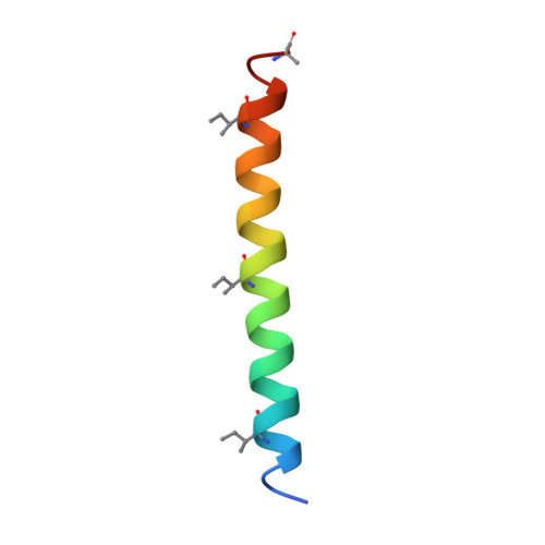
1RH4 - PubMed Abstract:
Recent advances in computational techniques have allowed the design of precise side-chain packing in proteins with predetermined, naturally occurring backbone structures. Because these methods do not model protein main-chain flexibility, they lack the breadth to explore novel backbone conformations. Here the de novo design of a family of alpha-helical bundle proteins with a right-handed superhelical twist is described. In the design, the overall protein fold was specified by hydrophobic-polar residue patterning, whereas the bundle oligomerization state, detailed main-chain conformation, and interior side-chain rotamers were engineered by computational enumerations of packing in alternate backbone structures. Main-chain flexibility was incorporated through an algebraic parameterization of the backbone. The designed peptides form alpha-helical dimers, trimers, and tetramers in accord with the design goals. The crystal structure of the tetramer matches the designed structure in atomic detail.
- Whitehead Institute for Biomedical Research, Howard Hughes Medical Institute and Department of Biology, Massachusetts Institute of Technology, Nine Cambridge Center, Cambridge, MA 02142, USA.
Organizational Affiliation: