Comparison of the structure and DNA-binding properties of the E2 proteins from an oncogenic and a non-oncogenic human papillomavirus.
Dell, G., Wilkinson, K.W., Tranter, R., Parish, J., Leo Brady, R., Gaston, K.(2003) J Mol Biology 334: 979-991
- PubMed: 14643661 Search on PubMed
- DOI: https://doi.org/10.1016/j.jmb.2003.10.009
- Primary Citation Related Structures:
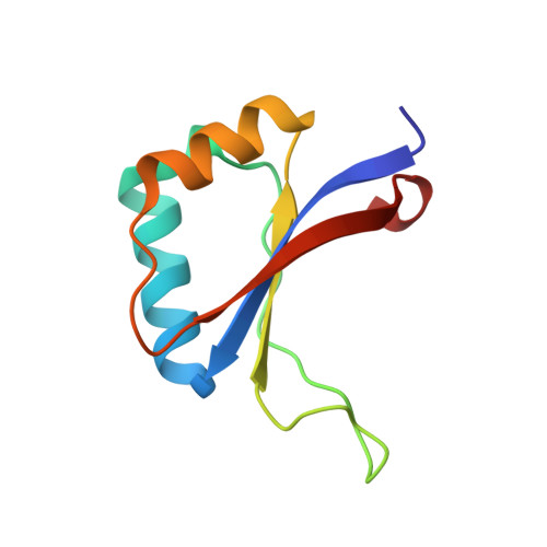
1R8H - PubMed Abstract:
Human papillomaviruses (HPVS) that infect the genital tract can be divided into two groups: high-risk HPV types, such as HPV 16 and HPV 18, are associated with cancer, low-risk HPV types, such as HPV 6, are associated with benign warts. In both high-risk and low-risk HPV types, the papillomavirus E2 protein binds to four sites within the viral long control region (LCR) and regulates viral gene expression. Here, we present the crystal structure of the minimal DNA-binding domain (DBD) from the HPV 6 E2 protein. We show that the HPV 6 E2 DBD is structurally more similar to the HPV 18 and bovine papillomavirus type 1 (BPV1) E2 proteins than it is to the HPV 16 E2 protein. Using gel retardation assays, we show that the hierarchy of E2 sites within the HPV 16 and HPV 6 LCRs are different. However, despite these differences in structure and site preference, both the HPV 16 and 6 E2 DBDs recognise an extended version of the consensus E2 binding site derived from studies of the BPV1 E2 protein. In both cases, the preferred binding site is 5'AACCGN(4)CGGTT3', where the additional flanking base-pairs are in bold and N(4) represents a four base-pair central spacer. Both of these HPV proteins bind preferentially to E2 sites that contain an A:T-rich central spacer. We show that the preference for an A:T-rich central spacer is due, at least in part, to the need to adopt a DNA conformation that facilitates protein contacts with the flanking base-pairs.
- Department of Biochemistry, School of Medical Sciences, University of Bristol, University Walk, Bristol BS8 1TD, UK.
Organizational Affiliation: