1QP5 | pdb_00001qp5

BASE-PAIRING SHIFT IN A DODECAMER CONTAINING A (CA)N TRACT


Experimental Data Snapshot

  • Method: X-RAY DIFFRACTION
  • Resolution: 2.60 Å
  • R-Value Observed: 
    0.161 (Depositor) 

wwPDB Validation 3D Report Full Report

Validation slider image for 1QP5

This is version 1.4 of the entry. See complete history

Literature

Base-pairing shift in the major groove of (CA)n tracts by B-DNA crystal structures.

Timsit, Y.Vilbois, E.Moras, D.

(1991) Nature 354: 167-170

  • DOI: https://doi.org/10.1038/354167a0
  • Primary Citation Related Structures: 
    1QP5, 330D

  • PubMed Abstract: 

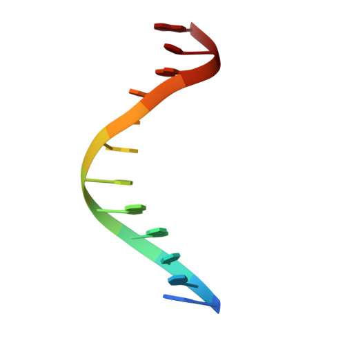
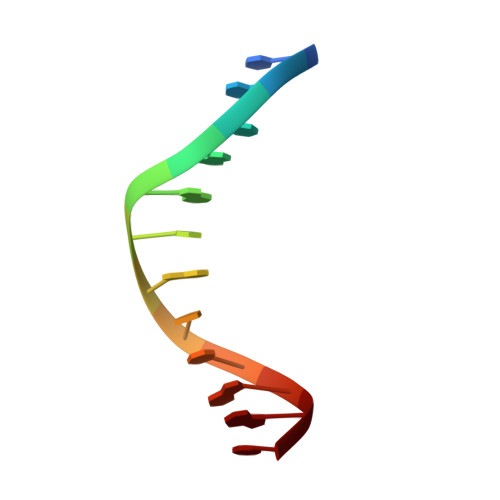
    the crystal packing of the B-DNA dodecamer d(ACCG-GCGCCACA).d(TGTGGCGCCGGT) is characterized by the reciprocal fit of double helices with specific base-backbone interactions in the major groove. Cooling the crystals below -10 degrees C stabilizes a new conformational state with a long-range sequence-dependent one-step shift in the major-groove base pairing. The tilt of the bases leads to the disruption of the Watson-Crick pairing in the major groove and to the formation of interactions with the 5' neighbour of their complement. This alteration propagates along the helical axis over more than half a turn. As a result, the molecular structure is normal when seen from the minor groove side and mismatched in the major groove. Comparison with a parent isomorphous dodecamer structure corresponding to the codon 10-13 of the c-Ha-ras proto-oncogene show that this new structural feature is sequence dependent and clearly favoured by (CA)n tracts. As(CA)n tracts of DNA are involved both in recombination and in transcription, this new recognition pattern should be considered in the analysis of the various processes involving the reading of the genetic information.


  • Organizational Affiliation
    • Laboratoire de Cristallographie Biologique, Institut de Biologie Moléculaire et Cellulaire, Strasbourg, France.

Macromolecule Content 

  • Total Structure Weight: 7.52 kDa 
  • Atom Count: 601 
  • Modeled Residue Count: 24 
  • Deposited Residue Count: 24 
  • Unique nucleic acid chains: 2

Macromolecules

Find similar nucleic acids by:  Sequence
Entity ID: 1
MoleculeChains LengthOrganismImage
DNA (5'-D(*AP*CP*CP*GP*GP*CP*GP*CP*CP*AP*CP*A)-3')12N/A
Sequence Annotations
Expand
Reference Sequence
Find similar nucleic acids by:  Sequence
Entity ID: 2
MoleculeChains LengthOrganismImage
DNA (5'-D(*TP*GP*TP*GP*GP*CP*GP*CP*CP*GP*GP*T)-3')12N/A
Sequence Annotations
Expand
Reference Sequence

Experimental Data & Validation

Experimental Data

  • Method: X-RAY DIFFRACTION
  • Resolution: 2.60 Å
  • R-Value Observed: 0.161 (Depositor) 
Space Group: H 3
Unit Cell:
Length ( Å )Angle ( ˚ )
a = 65.9α = 90
b = 65.9β = 90
c = 47.1γ = 120
Software Package:
Software NamePurpose
ULTIMAmodel building
NUCLSQrefinement
X-GENdata reduction
X-GENdata scaling
ULTIMAphasing

Structure Validation

View Full Validation Report


Entry History 

Deposition Data

Revision History  (Full details and data files)

  • Version 1.0: 1999-06-14
    Type: Initial release
  • Version 1.1: 2008-04-27
    Changes: Version format compliance
  • Version 1.2: 2011-07-13
    Changes: Version format compliance
  • Version 1.3: 2017-10-04
    Changes: Refinement description
  • Version 1.4: 2024-02-14
    Changes: Data collection, Database references, Derived calculations