Structure of Aspergillus Niger Epoxide Hydrolase at 1.8A Resolution: Implications for the Structure and Function of the Mammalian Microsomal Class of Epoxide Hydrolases
Zou, J.-Y., Hallberg, B.M., Bergfors, T., Oesch, F., Arand, M., Mowbray, S.L., Jones, T.A.(2000) Structure 8: 111
- PubMed: 10673439 Search on PubMed
- DOI: https://doi.org/10.1016/s0969-2126(00)00087-3
- Primary Citation Related Structures:
1QO7 - PubMed Abstract:
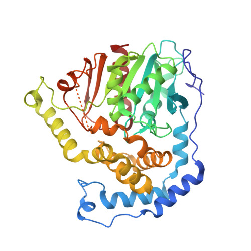
Epoxide hydrolases have important roles in the defense of cells against potentially harmful epoxides. Conversion of epoxides into less toxic and more easily excreted diols is a universally successful strategy. A number of microorganisms employ the same chemistry to process epoxides for use as carbon sources. The X-ray structure of the epoxide hydrolase from Aspergillus niger was determined at 3.5 A resolution using the multiwavelength anomalous dispersion (MAD) method, and then refined at 1.8 A resolution. There is a dimer consisting of two 44 kDa subunits in the asymmetric unit. Each subunit consists of an alpha/beta hydrolase fold, and a primarily helical lid over the active site. The dimer interface includes lid-lid interactions as well as contributions from an N-terminal meander. The active site contains a classical catalytic triad, and two tyrosines and a glutamic acid residue that are likely to assist in catalysis. The Aspergillus enzyme provides the first structure of an epoxide hydrolase with strong relationships to the most important enzyme of human epoxide metabolism, the microsomal epoxide hydrolase. Differences in active-site residues, especially in components that assist in epoxide ring opening and hydrolysis of the enzyme-substrate intermediate, might explain why the fungal enzyme attains the greater speeds necessary for an effective metabolic enzyme. The N-terminal domain that is characteristic of microsomal epoxide hydrolases corresponds to a meander that is critical for dimer formation in the Aspergillus enzyme.
- Department of Cell and Molecular Biology, BMC, Uppsala University, Box 596, Uppsala, S-751 24, Sweden.
Organizational Affiliation: