MAD Analyses of Yeast 5-Aminolaevulinic Acid Dehydratase. Their Use in Structure Determination and in Defining the Metal Binding Sites
Erskine, P.T., Duke, E.M.H., Tickle, I.J., Senior, N.M., Warren, M.J., Cooper, J.B.(2000) Acta Crystallogr D Biol Crystallogr 56: 421
- PubMed: 10739915 Search on PubMed
- DOI: https://doi.org/10.1107/s0907444900000597
- Primary Citation Related Structures:
1QML, 1QNV - PubMed Abstract:
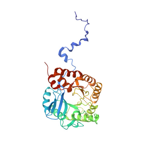
MAD experiments attempting to solve the structure of 5--aminolaevulinic acid dehydratase using Zn and Pb edges are described. The data obtained proved insufficient for a complete structure solution but were invaluable in subsequent identification of metal-binding sites using anomalous difference Fourier analyses once the structure of the enzyme had been solved. These sites include the highly inhibitory substitution of an enzymic cofactor Zn(2+) ion by Pb(2+) ions, which represents a major contribution towards understanding the molecular basis of lead poisoning. The MAD data collected at the Pb edge were also used with isomorphous replacement data from the same Pb co-crystal and a Hg co-crystal to provide the first delineation of the enzyme's quaternary structure. In this MADIR analysis, the Hg co-crystal data were treated as native data. Anomalous difference Fouriers were again used, revealing that Hg(2+) had substituted for the same Zn(2+) cofactor ion as had Pb(2+), a finding of fundamental importance for the understanding of mercury poisoning. In addition, Pt(2+) ions were found to bind at the same place in the structure. The refined structures of the Pb- and the Hg-complexed enzymes are presented at 2.5 and 3.0 A resolution, respectively.
- Biochemistry and Molecular Biology, School of Biological Sciences, University of Southampton, Bassett Crescent East, Southampton SO16 7PX, England.
Organizational Affiliation: