1PK8 | pdb_00001pk8

Crystal Structure of Rat Synapsin I C Domain Complexed to Ca.ATP


Experimental Data Snapshot

  • Method: X-RAY DIFFRACTION
  • Resolution: 2.10 Å
  • R-Value Free: 
    0.259 (Depositor), 0.267 (DCC) 
  • R-Value Work: 
    0.222 (Depositor), 0.232 (DCC) 
  • R-Value Observed: 
    0.222 (Depositor) 

Starting Model: experimental
View more details

wwPDB Validation 3D Report Full Report

Validation slider image for 1PK8

Ligand Structure Quality Assessment 


This is version 1.4 of the entry. See complete history

Literature

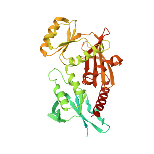
Tetramerization and ATP binding by a protein comprising the A, B, and C domains of rat synapsin I.

Brautigam, C.A.Chelliah, Y.Deisenhofer, J.

(2004) J Biological Chem 279: 11948-11956

  • DOI: https://doi.org/10.1074/jbc.M312015200
  • Primary Citation Related Structures: 
    1PK8, 1PX2

  • PubMed Abstract: 

    Synapsins are multidomain proteins that are critical for regulating neurotransmitter release in vertebrates. In the present study, two crystal structures of the C domain of rat synapsin I (rSynI-C) in complex with Ca(2+) and ATP reveal that this protein can form a tetramer and that a flexible loop (the "multifunctional loop") contacts bound ATP. Further experiments were carried out on a protein comprising the A, B, and C domains of rat synapsin I (rSynI-ABC). An ATP-stabilized tetramer of rSynI-ABC is observed during velocity sedimentation and size-exclusion chromatographic experiments. These hydrodynamic results also indicate that the A and B domains exist in an extended conformation. Calorimetric measurements of ATP binding to wild-type and mutant rSynI-ABC demonstrate that the multifunctional loop and a cross-tetramer contact are important for ATP binding. The evidence supports a view of synapsin I as an ATP-utilizing, tetrameric protein made up of monomers that have a flexible, extended N terminus.


  • Organizational Affiliation
    • Howard Hughes Medical Institute and the Department of Biochemistry, The University of Texas Southwestern Medical Center at Dallas, Dallas, Texas 75390-9050, USA.

Macromolecule Content 

  • Total Structure Weight: 368.82 kDa 
  • Atom Count: 19,708 
  • Modeled Residue Count: 2,334 
  • Deposited Residue Count: 3,376 
  • Unique protein chains: 1

Macromolecules

Find similar proteins by:|  3D Structure
Entity ID: 1
MoleculeChains  Sequence LengthOrganismDetailsImage
rat synapsin I
A, B, C, D, E
A, B, C, D, E, F, G, H
422Rattus norvegicusMutation(s): 0 
Gene Names: SYN1
UniProt
Find proteins for P09951 (Rattus norvegicus)
Explore P09951 
Go to UniProtKB:  P09951
Entity Groups
Sequence Clusters30% Identity50% Identity70% Identity90% Identity95% Identity100% Identity
UniProt GroupP09951
Sequence Annotations
Expand
Reference Sequence

Small Molecules

Ligands 3 Unique
IDChains Name / Formula / InChI Key2D Diagram3D Interactions
ATP

Query on ATP


Download:Ideal Coordinates CCD File
J [auth A]
M [auth B]
P [auth C]
R [auth D]
T [auth E]
J [auth A],
M [auth B],
P [auth C],
R [auth D],
T [auth E],
V [auth F],
X [auth G],
Z [auth H]
ADENOSINE-5'-TRIPHOSPHATE
C10 H16 N5 O13 P3
ZKHQWZAMYRWXGA-KQYNXXCUSA-N
EDO

Query on EDO


Download:Ideal Coordinates CCD File
K [auth A],
N [auth B]
1,2-ETHANEDIOL
C2 H6 O2
LYCAIKOWRPUZTN-UHFFFAOYSA-N
CA

Query on CA


Download:Ideal Coordinates CCD File
I [auth A]
L [auth B]
O [auth C]
Q [auth D]
S [auth E]
I [auth A],
L [auth B],
O [auth C],
Q [auth D],
S [auth E],
U [auth F],
W [auth G],
Y [auth H]
CALCIUM ION
Ca
BHPQYMZQTOCNFJ-UHFFFAOYSA-N

Experimental Data & Validation

Experimental Data

  • Method: X-RAY DIFFRACTION
  • Resolution: 2.10 Å
  • R-Value Free:  0.259 (Depositor), 0.267 (DCC) 
  • R-Value Work:  0.222 (Depositor), 0.232 (DCC) 
  • R-Value Observed: 0.222 (Depositor) 
Space Group: P 1
Unit Cell:
Length ( Å )Angle ( ˚ )
a = 70.6α = 80.6
b = 78.4β = 76.9
c = 135γ = 71.8
Software Package:
Software NamePurpose
HKL-2000data collection
SCALEPACKdata scaling
EPMRphasing
CNSrefinement
HKL-2000data reduction

Structure Validation

View Full Validation Report


Ligand Structure Quality Assessment 


Entry History 

Deposition Data

Revision History  (Full details and data files)

  • Version 1.0: 2004-03-23
    Type: Initial release
  • Version 1.1: 2008-04-29
    Changes: Version format compliance
  • Version 1.2: 2011-07-13
    Changes: Version format compliance
  • Version 1.3: 2023-08-16
    Changes: Data collection, Database references, Derived calculations, Refinement description
  • Version 1.4: 2024-03-13
    Changes: Source and taxonomy, Structure summary