1.67-A X-ray structure of the B2 immunoglobulin-binding domain of streptococcal protein G and comparison to the NMR structure of the B1 domain.
Achari, A., Hale, S.P., Howard, A.J., Clore, G.M., Gronenborn, A.M., Hardman, K.D., Whitlow, M.(1992) Biochemistry 31: 10449-10457
- PubMed: 1420164 Search on PubMed
- DOI: https://doi.org/10.1021/bi00158a006
- Primary Citation Related Structures:
1PGX - PubMed Abstract:
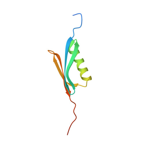
The structure of the B2 immunoglobulin-binding domain of streptococcal protein G has been determined at 1.67-A resolution using a combination of single isomorphous replacement (SIR) phasing and manual fitting of the coordinates of the NMR structure of B1 domain of streptococcal protein G [Gronenborn, A. M., et al. (1991) Science 253, 657-661]. The final R value was 0.191 for data between 8.0 and 1.67 A. The structure described here has 13 residues preceding the 57-residue Ig-binding domain and 13 additional residues following it, for a total of 83 residues. The 57-residue binding domain is well-determined in the structure, having an average B factor of 18.0. Only residues 8-77 could be located in the electron density maps, with the ends of the structure fading into disorder. Like the B1 domain, the B2 domain consists of four beta-strands and a single helix lying diagonally across the beta-sheet, with a -1, +3 chi, -1 topology. This small structure is extensively hydrogen-bonded and has a relatively large hydrophobic core. These structural observations may account for the exceptional stability of protein G. A comparison of the B2 domain X-ray structure and the B1 domain NMR structure showed minor differences in the turn between strands and two and a slight displacement of the helix relative to the sheet. Hydrogen bonds between crystallographically related molecules account for most of these differences.
- Protein Engineering Department, Enzon, Incorporated, Gaithersburg, Maryland 20877.
Organizational Affiliation: