1PG1 | pdb_00001pg1

PROTEGRIN 1 (PG1) FROM PORCINE LEUKOCYTES, NMR, 20 STRUCTURES


Experimental Data Snapshot

  • Method: SOLUTION NMR
  • Conformers Calculated: 100 
  • Conformers Submitted: 20 
  • Selection Criteria: LEAST RESTRAINT VIOLATION, LEAST GEOMETRY VIOLATION 

wwPDB Validation 3D Report Full Report

Validation slider image for 1PG1

This is version 1.4 of the entry. See complete history

Literature

Solution structure of protegrin-1, a broad-spectrum antimicrobial peptide from porcine leukocytes.

Fahrner, R.L.Dieckmann, T.Harwig, S.S.Lehrer, R.I.Eisenberg, D.Feigon, J.

(1996) Chem Biol 3: 543-550

  • DOI: https://doi.org/10.1016/s1074-5521(96)90145-3
  • Primary Citation Related Structures: 
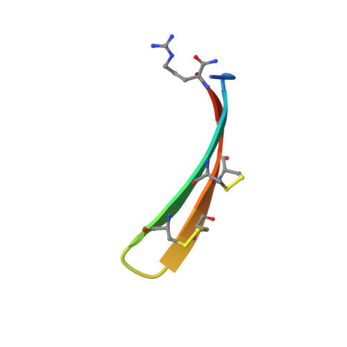
    1PG1

  • PubMed Abstract: 

    The protegrins are a family of arginine- and cysteine-rich cationic peptides found in porcine leukocytes that exhibit a broad range of antimicrobial and antiviral activities. They are composed of 16-18 amino-acid residues including four cysteines, which form two disulfide linkages. To begin to understand the mechanism of action of these peptides, we set out to determine the structure of protegrin-1 (PG-1). We used two-dimensional homonuclear nuclear magnetic resonance spectroscopy to study the conformation of both natural and synthetic PG-1 under several conditions. A refined three-dimensional structure of synthetic PG-1 is presented. Both synthetic and natural protegrin-1 form a well-defined structure in solution composed primarily of a two-stranded antiparallel beta sheet, with strands connected by a beta turn. The structure of PG-1 suggests ways in which the peptide may interact with itself or other molecules to form the membrane pores and the large membrane-associated assemblages observed in protegrin-treated, gram-negative bacteria.


  • Organizational Affiliation
    • Department of Chemistry and Biochemistry, University of California, Los Angeles, CA 90095, USA. feigon@ewald.mbi.ucla.edu

Macromolecule Content 

  • Total Structure Weight: 2.16 kDa 
  • Atom Count: 148 
  • Modeled Residue Count: 19 
  • Deposited Residue Count: 19 
  • Unique protein chains: 1

Macromolecules

Find similar proteins by:  Sequence   |   3D Structure  
Entity ID: 1
MoleculeChains  Sequence LengthOrganismDetailsImage
PROTEGRIN-119Sus scrofaMutation(s): 0 
Membrane Entity: Yes 
UniProt
Find proteins for P32194 (Sus scrofa)
Explore P32194 
Go to UniProtKB:  P32194
Entity Groups
Sequence Clusters30% Identity50% Identity70% Identity90% Identity95% Identity100% Identity
UniProt GroupP32194
Sequence Annotations
Expand
Reference Sequence

Experimental Data & Validation

Experimental Data

  • Method: SOLUTION NMR
  • Conformers Calculated: 100 
  • Conformers Submitted: 20 
  • Selection Criteria: LEAST RESTRAINT VIOLATION, LEAST GEOMETRY VIOLATION 

Structure Validation

View Full Validation Report


Entry History 

Deposition Data

Revision History  (Full details and data files)

  • Version 1.0: 1998-05-27
    Type: Initial release
  • Version 1.1: 2008-03-24
    Changes: Version format compliance
  • Version 1.2: 2011-07-13
    Changes: Version format compliance
  • Version 1.3: 2022-02-23
    Changes: Database references, Derived calculations, Other
  • Version 1.4: 2024-11-20
    Changes: Data collection, Structure summary