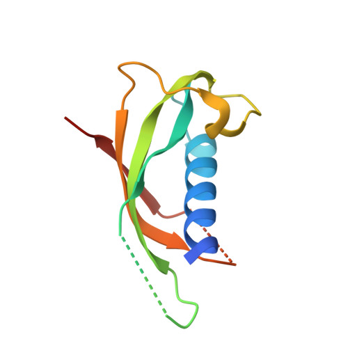
1PFP

CATHELIN-LIKE MOTIF OF PROTEGRIN-3


Experimental Data Snapshot

  • Method: X-RAY DIFFRACTION
  • Resolution: 2.30 Å
  • R-Value Free: 0.284 
  • R-Value Work: 0.219 
  • R-Value Observed: 0.227 

wwPDB Validation   3D Report Full Report


This is version 1.5 of the entry. See complete history


Literature

Selenomethionine and Selenocysteine Double Labeling Strategy for Crystallographic Phasing

Strub, M.-P.Hoh, F.Sanchez, J.-F.Strub, J.M.Bock, A.Aumelas, A.Dumas, C.

(2003) Structure 11: 1359-1367

  • DOI: https://doi.org/10.1016/j.str.2003.09.014
  • Primary Citation of Related Structures:  
    1PAE, 1PFP

  • PubMed Abstract: 

    A protocol for the quantitative incorporation of both selenomethionine and selenocysteine into recombinant proteins overexpressed in Escherichia coli is described. This methodology is based on the use of a suitable cysteine auxotrophic strain and a minimal medium supplemented with selenium-labeled methionine and cysteine. The proteins chosen for these studies are the cathelin-like motif of protegrin-3 and a nucleoside-diphosphate kinase. Analysis of the purified proteins by electrospray mass spectrometry and X-ray crystallography revealed that both cysteine and methionine residues were isomorphously replaced by selenocysteine and selenomethionine. Moreover, selenocysteines allowed the formation of unstrained and stable diselenide bridges in place of the canonical disulfide bonds. In addition, we showed that NDP kinase contains a selenocysteine adduct on Cys122. This novel selenium double-labeling method is proposed as a general approach to increase the efficiency of the MAD technique used for phase determination in protein crystallography.


  • Organizational Affiliation

    Centre de Biochimie Structurale, UMR CNRS 5048, UMR 554 INSERM, Université Montpellier I, 34090 Cedex, Montpellier, France.


Macromolecules
Find similar proteins by:  (by identity cutoff)  |  3D Structure
Entity ID: 1
MoleculeChains Sequence LengthOrganismDetailsImage
Protegrin 3105Sus scrofaMutation(s): 4 
Gene Names: NPG3
UniProt
Find proteins for P32196 (Sus scrofa)
Explore P32196 
Go to UniProtKB:  P32196
Entity Groups  
Sequence Clusters30% Identity50% Identity70% Identity90% Identity95% Identity100% Identity
UniProt GroupP32196
Sequence Annotations
Expand
  • Reference Sequence
Small Molecules
Modified Residues  1 Unique
IDChains TypeFormula2D DiagramParent
MSE
Query on MSE
A
L-PEPTIDE LINKINGC5 H11 N O2 SeMET
Experimental Data & Validation

Experimental Data

  • Method: X-RAY DIFFRACTION
  • Resolution: 2.30 Å
  • R-Value Free: 0.284 
  • R-Value Work: 0.219 
  • R-Value Observed: 0.227 
  • Space Group: P 65 2 2
Unit Cell:
Length ( Å )Angle ( ˚ )
a = 51.541α = 90
b = 51.541β = 90
c = 134.512γ = 120
Software Package:
Software NamePurpose
REFMACrefinement
DENZOdata reduction
SCALEPACKdata scaling
SHARPphasing

Structure Validation

View Full Validation Report


Entry History 

Deposition Data

Revision History  (Full details and data files)

  • Version 1.0: 2003-11-11
    Type: Initial release
  • Version 1.1: 2008-04-29
    Changes: Version format compliance
  • Version 1.2: 2011-07-13
    Changes: Version format compliance
  • Version 1.3: 2014-02-19
    Changes: Atomic model, Structure summary
  • Version 1.4: 2014-08-06
    Changes: Derived calculations, Structure summary
  • Version 1.5: 2024-02-14
    Changes: Data collection, Database references