Crystal structure of the complex of phosphofructokinase from Escherichia coli with its reaction products.
Shirakihara, Y., Evans, P.R.(1988) J Mol Biology 204: 973-994
- PubMed: 2975709 Search on PubMed
- DOI: https://doi.org/10.1016/0022-2836(88)90056-3
- Primary Citation Related Structures:
1PFK - PubMed Abstract:
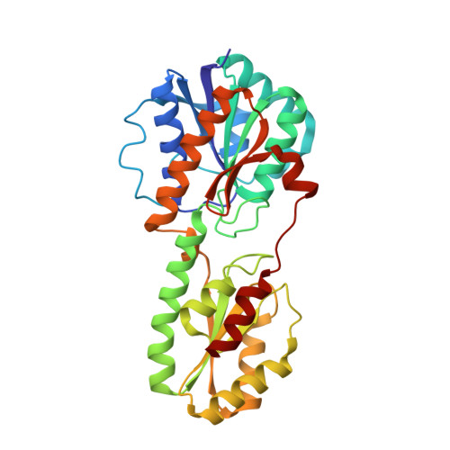
The crystal structure of Escherichia coli phosphofructokinase complexed with its reaction products fructose 1,6-bisphosphate (Fru1,6P) and ADP/Mg2+, and the allosteric activator ADP/Mg2+, has been determined at 2.4 A resolution. The structure was solved by molecular replacement using the known structure of Bacillus stearothermophilus phosphofructokinase, and has been refined to a crystallographic R-factor of 0.165 for all data. The crystallization mixture contained the substrate fructose 6-phosphate, but the electron density maps showed clearly the presence of the product fructose 1,6-bisphosphate, presumably formed by the enzyme reaction with contaminating ATP. The crystal consists of tetrameric molecules with subunits in two different conformations despite their chemical identity. The magnesium ion in the "closed" subunit bridges the phosphate groups of the two products. In the "open" subunit, the products are about 1.5 A further apart, with the Mg2+ bound only to ADP. These two conformations probably represent two successive stages along the reaction pathway, in which the closure of the subunit is required to bring the substrates sufficiently close to react. This conformational change within the subunit is distinct from the quaternary structure change seen previously in the inactive T-state conformation. It is probably not involved in the co-operativity or allosteric control of the enzyme, since the co-operative product fructose 1,6-bisphosphate is not moved, nor are the subunit interfaces changed. The structure of the enzyme is similar to that of B. stearothermophilus phosphofructokinase, and confirms the location of the sites for the two reaction products (or substrates), and of the effector site binding the activator ADP/Mg2+. However, this structure gives a clearer picture of the active site, and of the interactions between the enzyme and its reaction products.
- Laboratory of Molecular Biology, Cambridge, England.
Organizational Affiliation: