Response of a designed metalloprotein to changes in metal ion coordination, exogenous ligands, and active site volume determined by X-ray crystallography.
Geremia, S., Di Costanzo, L., Randaccio, L., Engel, D.E., Lombardi, A., Nastri, F., DeGrado, W.F.(2005) J Am Chem Soc 127: 17266-17276
- PubMed: 16332076 Search on PubMed
- DOI: https://doi.org/10.1021/ja054199x
- Primary Citation Related Structures:
1OVR, 1OVU, 1OVV - PubMed Abstract:
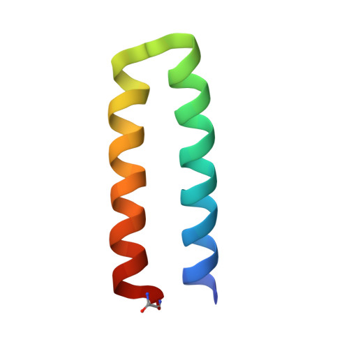
The de novo protein DF1 is a minimal model for diiron and dimanganese metalloproteins, such as soluble methane monooxygenase. DF1 is a homodimeric four-helix bundle whose dinuclear center is formed by two bridging Glu side chains, two chelating Glu side chains, and two monodentate His ligands. Here, we report the di-Mn(II) and di-Co(II) derivatives of variants of this protein. Together with previously solved structures, 23 crystallographically independent four-helix bundle structures of DF1 variants have been determined, which differ in the bound metal ions and size of the active site cavity. For the di-Mn(II) derivatives, as the size of the cavity increases, the number and polarity of exogenous ligands increases. This collection of structures was analyzed to determine the relationship between protein conformation and the geometry of the active site. The primary mode of backbone movement involves a coordinated tilting and sliding of the first helix in the helix-loop-helix motif. Sliding depends on crystal-packing forces, the steric bulk of a critical residue that determines the dimensions of the active site access cavity, and the intermetal distance. Additionally, a torsional motion of the bridging carboxylates modulates the intermetal distance. This analysis provides a critical evaluation of how conformation, flexibility, and active site accessibility affect the geometry and ligand-binding properties of a metal center. The geometric parameters defining the DF structures were compared to natural diiron proteins; DF proteins have a restricted active site cavity, which may have implications for substrate recognition and chemical stability.
- Centre of Excellence in Biocrystallography, Department of Chemical Science, University of Trieste, Via L. Giorgieri 1, I-34127 Trieste, Italy. geremia@units.it
Organizational Affiliation: