1OGU | pdb_00001ogu

STRUCTURE OF HUMAN THR160-PHOSPHO CDK2/CYCLIN A COMPLEXED WITH A 2-ARYLAMINO-4-CYCLOHEXYLMETHYL-5-NITROSO-6-AMINOPYRIMIDINE INHIBITOR


Experimental Data Snapshot

  • Method: X-RAY DIFFRACTION
  • Resolution: 2.60 Å
  • R-Value Free: 
    0.260 (Depositor), 0.254 (DCC) 
  • R-Value Work: 
    0.198 (Depositor), 0.194 (DCC) 
  • R-Value Observed: 
    0.201 (Depositor) 

Starting Model: experimental
View more details

wwPDB Validation 3D Report Full Report

Validation slider image for 1OGU

Ligand Structure Quality Assessment 


This is version 1.4 of the entry. See complete history

Literature

Structure-Based Design of 2-Arylamino-4-Cyclohexyl Methyl-5-Nitroso-6-Aminopyrimidine Inhibitors of Cyclin-Dependent Kinases 1 and 2

Sayle, K.L.Bentley, J.Boyle, F.T.Calvert, A.H.Cheng, Y.Curtin, N.J.Endicott, J.A.Golding, B.T.Hardcastle, I.R.Jewsbury, P.Mesguiche, V.Newell, D.R.Noble, M.E.M.Parsons, R.J.Pratt, D.J.Wang, L.Z.Griffin, R.J.

(2003) Bioorg Med Chem Lett 13: 3079

  • DOI: https://doi.org/10.1016/s0960-894x(03)00651-6
  • Primary Citation Related Structures: 
    1OGU

  • PubMed Abstract: 

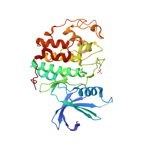
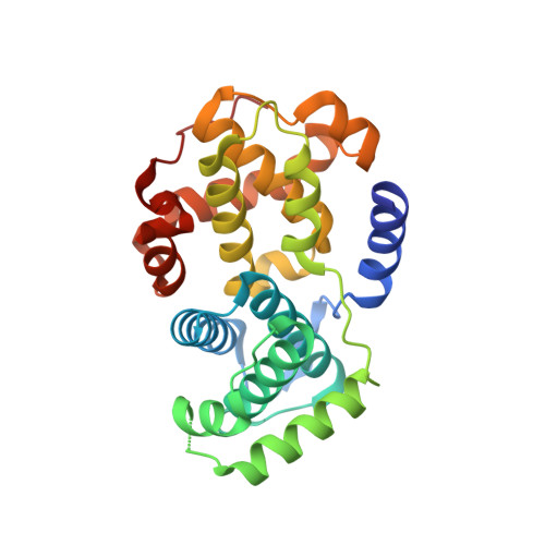
    A series of O(4)-cyclohexylmethyl-5-nitroso-6-aminopyrimidines bearing 2-arylamino substituents was synthesised and evaluated for CDK1 and CDK2 inhibitory activity. Consistent with analogous studies with O(6)-cyclohexylmethylpurines, 2-arylaminopyrimidines with a sulfonamide or carboxamide group at the 4'-position were potent inhibitors, with IC(50) values against CDK2 of 1.1+/-0.3 and 34+/-8 nM, respectively. The crystal structure of the 4'-carboxamide derivative, in complex with phospho-Thr160 CDK2/cyclin A, confirmed the expected binding mode of the inhibitor, and revealed an additional interaction between the carboxamide function and an aspartate residue.


  • Organizational Affiliation
    • Northern Institute for Cancer Research, School of Natural Sciences-Chemistry, Bedson Building, University of Newcastle, Newcastle Upon Tyne NE1 7RU, UK.

Macromolecule Content 

  • Total Structure Weight: 129.44 kDa 
  • Atom Count: 8,893 
  • Modeled Residue Count: 1,078 
  • Deposited Residue Count: 1,124 
  • Unique protein chains: 2

Macromolecules

Find similar proteins by:|  3D Structure
Entity ID: 1
MoleculeChains  Sequence LengthOrganismDetailsImage
CELL DIVISION PROTEIN KINASE 2
A, C
302Homo sapiensMutation(s): 0 
EC: 2.7.1.37 (PDB Primary Data), 2.7.11.22 (UniProt)
UniProt & NIH Common Fund Data Resources
Find proteins for P24941 (Homo sapiens)
Explore P24941 
Go to UniProtKB:  P24941
PHAROS:  P24941
GTEx:  ENSG00000123374 
Entity Groups
Sequence Clusters30% Identity50% Identity70% Identity90% Identity95% Identity100% Identity
UniProt GroupP24941
Sequence Annotations
Expand
Reference Sequence
Find similar proteins by:|  3D Structure
Entity ID: 2
MoleculeChains  Sequence LengthOrganismDetailsImage
CYCLIN A2
B, D
260Homo sapiensMutation(s): 0 
UniProt & NIH Common Fund Data Resources
Find proteins for P20248 (Homo sapiens)
Explore P20248 
Go to UniProtKB:  P20248
PHAROS:  P20248
GTEx:  ENSG00000145386 
Entity Groups
Sequence Clusters30% Identity50% Identity70% Identity90% Identity95% Identity100% Identity
UniProt GroupP20248
Sequence Annotations
Expand
Reference Sequence

Small Molecules

Modified Residues  1 Unique
IDChains TypeFormula2D DiagramParent
TPO
Query on TPO
A, C
L-PEPTIDE LINKINGC4 H10 N O6 PTHR
Binding Affinity Annotations 
IDSourceBinding Affinity
ST8 BindingDB:  1OGU IC50: 34 (nM) from 1 assay(s)

Experimental Data & Validation

Experimental Data

  • Method: X-RAY DIFFRACTION
  • Resolution: 2.60 Å
  • R-Value Free:  0.260 (Depositor), 0.254 (DCC) 
  • R-Value Work:  0.198 (Depositor), 0.194 (DCC) 
  • R-Value Observed: 0.201 (Depositor) 
Space Group: P 21 21 21
Unit Cell:
Length ( Å )Angle ( ˚ )
a = 73.836α = 90
b = 133.676β = 90
c = 148.335γ = 90
Software Package:
Software NamePurpose
REFMACrefinement
MOSFLMdata reduction
SCALAdata scaling

Structure Validation

View Full Validation Report


Ligand Structure Quality Assessment 


Entry History 

Deposition Data

Revision History  (Full details and data files)

  • Version 1.0: 2003-09-02
    Type: Initial release
  • Version 1.1: 2011-05-08
    Changes: Version format compliance
  • Version 1.2: 2011-07-13
    Changes: Version format compliance
  • Version 1.3: 2023-12-13
    Changes: Data collection, Database references, Derived calculations, Other, Refinement description
  • Version 1.4: 2024-11-06
    Changes: Structure summary