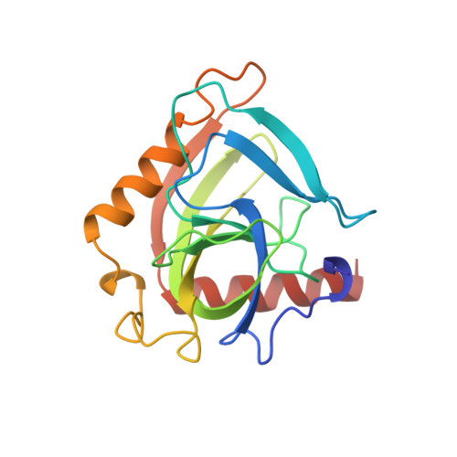
1OBW

STRUCTURE OF INORGANIC PYROPHOSPHATASE


Experimental Data Snapshot

  • Method: X-RAY DIFFRACTION
  • Resolution: 1.90 Å
  • R-Value Free: 0.220 
  • R-Value Work: 0.176 

Starting Model: experimental
View more details

wwPDB Validation   3D Report Full Report


This is version 1.4 of the entry. See complete history


Literature

Crystal structure of holo inorganic pyrophosphatase from Escherichia coli at 1.9 A resolution. Mechanism of hydrolysis.

Harutyunyan, E.H.Oganessyan, V.Y.Oganessyan, N.N.Avaeva, S.M.Nazarova, T.I.Vorobyeva, N.N.Kurilova, S.A.Huber, R.Mather, T.

(1997) Biochemistry 36: 7754-7760

  • DOI: https://doi.org/10.1021/bi962637u
  • Primary Citation of Related Structures:  
    1OBW

  • PubMed Abstract: 

    Crystalline holo inorganic pyrophosphatase from Escherichia coli was grown in the presence of 250 mM MgCl2. The crystal structure has been solved by Patterson search techniques and refined to an R-factor of 17.6% at 1.9 A resolution. The upper estimate of the root-mean-square error in atomic positions is 0.26 A. These crystals belong to space group P3(2)21 with unit cell dimensions a = b = 110.27 A and c = 78.17 A. The asymmetric unit contains a trimer of subunits, i.e., half of the hexameric molecule. In the central cavity of the enzyme molecule, three Mg2+ ions, each shared by two subunits of the hexamer, are found. In the active sites of two crystallographically independent subunits, two Mg2+ ions are bound. The second active site Mg2+ ion is missing in the third subunit. A mechanism of catalysis is proposed whereby a water molecule activated by a Mg2+ ion and Tyr 55 play essential roles.


  • Organizational Affiliation

    Shubnikov Institute of Crystallography, Russian Academy of Sciences, Moscow, Russian Federation. emil@protein.crystal.msk.ru


Macromolecules
Find similar proteins by:  (by identity cutoff)  |  3D Structure
Entity ID: 1
MoleculeChains Sequence LengthOrganismDetailsImage
INORGANIC PYROPHOSPHATASE
A, B, C
175Escherichia coliMutation(s): 0 
EC: 3.6.1.1
UniProt
Find proteins for P0A7A9 (Escherichia coli (strain K12))
Explore P0A7A9 
Go to UniProtKB:  P0A7A9
Entity Groups  
Sequence Clusters30% Identity50% Identity70% Identity90% Identity95% Identity100% Identity
UniProt GroupP0A7A9
Sequence Annotations
Expand
  • Reference Sequence
Experimental Data & Validation

Experimental Data

  • Method: X-RAY DIFFRACTION
  • Resolution: 1.90 Å
  • R-Value Free: 0.220 
  • R-Value Work: 0.176 
  • Space Group: P 32 2 1
Unit Cell:
Length ( Å )Angle ( ˚ )
a = 110.3α = 90
b = 110.3β = 90
c = 78.2γ = 120
Software Package:
Software NamePurpose
AMoREphasing
REFMACrefinement
DENZOdata reduction
SCALEPACKdata scaling

Structure Validation

View Full Validation Report


Entry History 

Deposition Data

Revision History  (Full details and data files)

  • Version 1.0: 1997-09-04
    Type: Initial release
  • Version 1.1: 2008-03-24
    Changes: Version format compliance
  • Version 1.2: 2011-07-13
    Changes: Derived calculations, Version format compliance
  • Version 1.3: 2024-02-14
    Changes: Data collection, Database references, Derived calculations
  • Version 1.4: 2024-04-03
    Changes: Refinement description