1OBW

STRUCTURE OF INORGANIC PYROPHOSPHATASE


Experimental Data Snapshot

  • Method: X-RAY DIFFRACTION
  • Resolution: 1.90 Å
  • R-Value Free: 0.220 
  • R-Value Work: 0.176 

wwPDB Validation   3D Report Full Report


This is version 1.4 of the entry. See complete history


Literature

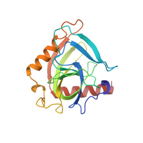
Crystal structure of holo inorganic pyrophosphatase from Escherichia coli at 1.9 A resolution. Mechanism of hydrolysis.

Harutyunyan, E.H.Oganessyan, V.Y.Oganessyan, N.N.Avaeva, S.M.Nazarova, T.I.Vorobyeva, N.N.Kurilova, S.A.Huber, R.Mather, T.

(1997) Biochemistry 36: 7754-7760

  • DOI: https://doi.org/10.1021/bi962637u
  • Primary Citation of Related Structures:  
    1OBW

  • PubMed Abstract: 

    Crystalline holo inorganic pyrophosphatase from Escherichia coli was grown in the presence of 250 mM MgCl2. The crystal structure has been solved by Patterson search techniques and refined to an R-factor of 17.6% at 1.9 A resolution. The upper estimate of the root-mean-square error in atomic positions is 0.26 A. These crystals belong to space group P3(2)21 with unit cell dimensions a = b = 110.27 A and c = 78.17 A. The asymmetric unit contains a trimer of subunits, i.e., half of the hexameric molecule. In the central cavity of the enzyme molecule, three Mg2+ ions, each shared by two subunits of the hexamer, are found. In the active sites of two crystallographically independent subunits, two Mg2+ ions are bound. The second active site Mg2+ ion is missing in the third subunit. A mechanism of catalysis is proposed whereby a water molecule activated by a Mg2+ ion and Tyr 55 play essential roles.


  • Organizational Affiliation

    Shubnikov Institute of Crystallography, Russian Academy of Sciences, Moscow, Russian Federation. emil@protein.crystal.msk.ru


Macromolecules
Find similar proteins by:  (by identity cutoff)  |  3D Structure
Entity ID: 1
MoleculeChains Sequence LengthOrganismDetailsImage
INORGANIC PYROPHOSPHATASE
A, B, C
175Escherichia coliMutation(s): 0 
EC: 3.6.1.1
UniProt
Find proteins for P0A7A9 (Escherichia coli (strain K12))
Explore P0A7A9 
Go to UniProtKB:  P0A7A9
Entity Groups  
Sequence Clusters30% Identity50% Identity70% Identity90% Identity95% Identity100% Identity
UniProt GroupP0A7A9
Sequence Annotations
Expand
  • Reference Sequence
Experimental Data & Validation

Experimental Data

  • Method: X-RAY DIFFRACTION
  • Resolution: 1.90 Å
  • R-Value Free: 0.220 
  • R-Value Work: 0.176 
  • Space Group: P 32 2 1
Unit Cell:
Length ( Å )Angle ( ˚ )
a = 110.3α = 90
b = 110.3β = 90
c = 78.2γ = 120
Software Package:
Software NamePurpose
AMoREphasing
REFMACrefinement
DENZOdata reduction
SCALEPACKdata scaling

Structure Validation

View Full Validation Report


Entry History 

Deposition Data

Revision History  (Full details and data files)

  • Version 1.0: 1997-09-04
    Type: Initial release
  • Version 1.1: 2008-03-24
    Changes: Version format compliance
  • Version 1.2: 2011-07-13
    Changes: Derived calculations, Version format compliance
  • Version 1.3: 2024-02-14
    Changes: Data collection, Database references, Derived calculations
  • Version 1.4: 2024-04-03
    Changes: Refinement description