1O8J

Pectate Lyase C from Erwinia Chrysanthemi at pH 4.5 with 30mM CA2+


Experimental Data Snapshot

  • Method: X-RAY DIFFRACTION
  • Resolution: 2.20 Å
  • R-Value Free: 0.222 
  • R-Value Work: 0.176 
  • R-Value Observed: 0.176 

wwPDB Validation   3D Report Full Report


This is version 2.1 of the entry. See complete history


Literature

Characterization and Implications of Ca2+ Binding to Pectate Lyase C

Herron, S.R.Scavetta, R.Garrett, M.Legner, M.Jurnak, F.A.

(2003) J Biol Chem 278: 12271-12277

  • DOI: https://doi.org/10.1074/jbc.M209306200
  • Primary Citation of Related Structures:  
    1O88, 1O8D, 1O8E, 1O8F, 1O8G, 1O8H, 1O8I, 1O8J, 1O8K, 1O8L, 1O8M

  • PubMed Abstract: 

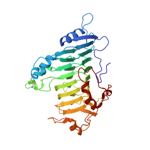
    Ca(2+) is essential for in vitro activity of Erwinia chrysanthemi pectate lyase C (PelC). Crystallographic analyses of 11 PelC-Ca(2+) complexes, formed at pH 4.5, 9.5, and 11.2 under varying Ca(2+) concentrations, have been solved and refined at a resolution of 2.2 A. The Ca(2+) site represents a new motif for Ca(2+), consisting primarily of beta-turns and beta-strands. The principal differences between PelC and the PelC-Ca(2+) structures at all pH values are the side-chain conformations of Asp-129 and Glu-166 as well as the occupancies of four water molecules. According to calculations of pK(a) values, the presence of Ca(2+) and associated structural changes lower the pK(a) of Arg-218, the amino acid responsible for proton abstraction during catalysis. The Ca(2+) affinity for PelC is weak, as the K(d) was estimated to be 0.132 (+/-0.004) mm at pH 9.5, 1.09 (+/-0.29) mm at pH 11.2, and 5.84 (+/-0.41) mm at pH 4.5 from x-ray diffraction studies and 0.133 (+/-0.045) mm at pH 9.5 from intrinsic tryptophan fluorescence measurements. Given the pH dependence of Ca(2+) affinity, PelC activity at pH 4.5 has been reexamined. At saturating Ca(2+) concentrations, PelC activity increases 10-fold at pH 4.5 but is less than 1% of maximal activity at pH 9.5. Taken together, the studies suggest that the primary Ca(2+) ion in PelC has multiple functions.


  • Organizational Affiliation

    Department of Physiology and Biophysics, University of California, Irvine, California 92697-4560, USA.


Macromolecules
Find similar proteins by:  (by identity cutoff)  |  3D Structure
Entity ID: 1
MoleculeChains Sequence LengthOrganismDetailsImage
PECTATE LYASE C353Dickeya chrysanthemiMutation(s): 0 
EC: 4.2.2.2
UniProt
Find proteins for P11073 (Dickeya chrysanthemi)
Explore P11073 
Go to UniProtKB:  P11073
Entity Groups  
Sequence Clusters30% Identity50% Identity70% Identity90% Identity95% Identity100% Identity
UniProt GroupP11073
Sequence Annotations
Expand
  • Reference Sequence
Small Molecules
Ligands 1 Unique
IDChains Name / Formula / InChI Key2D Diagram3D Interactions
CA
Query on CA

Download Ideal Coordinates CCD File 
B [auth A]CALCIUM ION
Ca
BHPQYMZQTOCNFJ-UHFFFAOYSA-N
Experimental Data & Validation

Experimental Data

  • Method: X-RAY DIFFRACTION
  • Resolution: 2.20 Å
  • R-Value Free: 0.222 
  • R-Value Work: 0.176 
  • R-Value Observed: 0.176 
  • Space Group: P 21 21 21
Unit Cell:
Length ( Å )Angle ( ˚ )
a = 72.89α = 90
b = 80.93β = 90
c = 95.83γ = 90
Software Package:
Software NamePurpose
X-PLORrefinement
SDMSdata reduction
SDMSdata scaling
X-PLORphasing

Structure Validation

View Full Validation Report


Entry History 

Deposition Data

Revision History  (Full details and data files)

  • Version 1.0: 2003-01-30
    Type: Initial release
  • Version 1.1: 2011-05-08
    Changes: Version format compliance
  • Version 1.2: 2011-07-13
    Changes: Version format compliance
  • Version 2.0: 2018-06-13
    Changes: Atomic model, Data collection, Database references, Structure summary
  • Version 2.1: 2023-12-13
    Changes: Data collection, Database references, Derived calculations, Other, Refinement description