1O75

Tp47, the 47-Kilodalton Lipoprotein of Treponema pallidum


Experimental Data Snapshot

  • Method: X-RAY DIFFRACTION
  • Resolution: 1.95 Å
  • R-Value Free: 0.228 
  • R-Value Work: 0.205 
  • R-Value Observed: 0.205 

wwPDB Validation   3D Report Full Report


This is version 2.0 of the entry. See complete history


Literature

Crystal structure of the 47-kDa lipoprotein of Treponema pallidum reveals a novel penicillin-binding protein.

Deka, R.K.Machius, M.Norgard, M.V.Tomchick, D.R.

(2002) J Biol Chem 277: 41857-41864

  • DOI: https://doi.org/10.1074/jbc.M207402200
  • Primary Citation of Related Structures:  
    1O75

  • PubMed Abstract: 

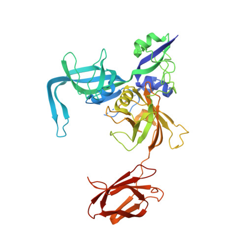
    Syphilis is a complex sexually transmitted disease caused by the spirochetal bacterium Treponema pallidum. T. pallidum has remained exquisitely sensitive to penicillin, but the mode of action and lethal targets for beta-lactams are still unknown. We previously identified the T. pallidum 47-kDa lipoprotein (Tp47) as a penicillin-binding protein (PBP). Tp47 contains three hypothetical consensus motifs (SVTK, TEN, and KTG) that typically form the active center of other PBPs. Yet, in this study, mutations of key amino acids within these motifs failed to abolish the penicillin binding activity of Tp47. The crystal structure of Tp47 at a resolution of 1.95 A revealed a fold different from any other known PBP; Tp47 is predominantly beta-sheet, in contrast to the alpha/beta-fold common to other PBPs. It comprises four distinct domains: two complex beta-sheet-containing N-terminal domains and two C-terminal domains that adopt immunoglobulin-like folds. The three hypothetical PBP signature motifs do not come together to form a typical PBP active site. Furthermore, Tp47 is unusual in that it displays beta-lactamase activity (k(cat) for penicillin = 271 +/- 6 s(-1)), a feature that hindered attempts to identify the active site in Tp47 by co-crystallization and mass spectrometric techniques. Taken together, Tp47 does not fit the classical structural and mechanistic paradigms for PBPs, and thus Tp47 appears to represent a new class of PBP.


  • Organizational Affiliation

    Departments of Microbiology and Biochemistry, University of Texas Southwestern Medical Center, Dallas 75390, USA.


Macromolecules
Find similar proteins by:  (by identity cutoff)  |  3D Structure
Entity ID: 1
MoleculeChains Sequence LengthOrganismDetailsImage
47 KDA MEMBRANE ANTIGEN
A, B
415Treponema pallidumMutation(s): 2 
UniProt
Find proteins for P29723 (Treponema pallidum (strain Nichols))
Explore P29723 
Go to UniProtKB:  P29723
Entity Groups  
Sequence Clusters30% Identity50% Identity70% Identity90% Identity95% Identity100% Identity
UniProt GroupP29723
Sequence Annotations
Expand
  • Reference Sequence
Oligosaccharides

Help

Entity ID: 2
MoleculeChains Length2D Diagram Glycosylation3D Interactions
2,3-di-O-sulfo-alpha-D-glucopyranose-(1-6)-2,3-di-O-sulfo-alpha-D-glucopyranose
C
2N/A
Glycosylation Resources
GlyTouCan:  G59221CU
GlyCosmos:  G59221CU
Experimental Data & Validation

Experimental Data

  • Method: X-RAY DIFFRACTION
  • Resolution: 1.95 Å
  • R-Value Free: 0.228 
  • R-Value Work: 0.205 
  • R-Value Observed: 0.205 
  • Space Group: P 32 2 1
Unit Cell:
Length ( Å )Angle ( ˚ )
a = 128.931α = 90
b = 128.931β = 90
c = 151.161γ = 120
Software Package:
Software NamePurpose
CNSrefinement
HKL-2000data reduction
SCALEPACKdata scaling
MLPHAREphasing

Structure Validation

View Full Validation Report


Entry History 

Deposition Data

Revision History  (Full details and data files)

  • Version 1.0: 2002-11-01
    Type: Initial release
  • Version 1.1: 2011-05-08
    Changes: Version format compliance
  • Version 1.2: 2011-07-13
    Changes: Version format compliance
  • Version 1.3: 2018-02-28
    Changes: Database references, Source and taxonomy
  • Version 2.0: 2020-07-29
    Type: Remediation
    Reason: Carbohydrate remediation
    Changes: Atomic model, Data collection, Derived calculations, Other, Structure summary