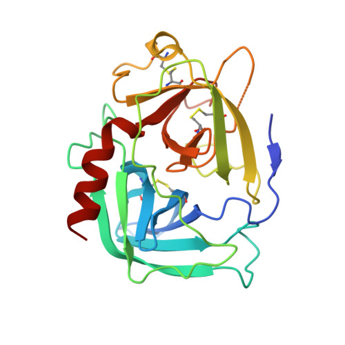
Structure of human pro-chymase: a model for the activating transition of granule-associated proteases.
Reiling, K.K., Krucinski, J., Miercke, L.J., Raymond, W.W., Caughey, G.H., Stroud, R.M.(2003) Biochemistry 42: 2616-2624
- PubMed: 12614156
- DOI: https://doi.org/10.1021/bi020594d
- Primary Citation of Related Structures:
1NN6 - PubMed Abstract:
Human chymase is a protease involved in physiological processes ranging from inflammation to hypertension. As are all proteases of the trypsin fold, chymase is synthesized as an inactive "zymogen" with an N-terminal pro region that prevents the transition of the zymogen to an activated conformation. The 1.8 A structure of pro-chymase, reported here, is the first zymogen with a dipeptide pro region (glycine-glutamate) to be characterized at atomic resolution. Three segments of the pro-chymase structure differ from that of the activated enzyme: the N-terminus (Gly14-Gly19), the autolysis loop (Gly142-Thr154), and the 180s loop (Pro185A-Asp194). The four N-terminal residues (Gly14-Glu15-Ile16-Ile17) are disordered. The autolysis loop occupies a position up to 10 A closer to the active site than is seen in the activated enzyme, thereby forming a hydrogen bond with the catalytic residue Ser195 and occluding the S1' binding pocket. Nevertheless, the catalytic triad (Asp102-His57-Ser195) is arrayed in a geometry close to that seen in activated chymase (all atom rmsd of 0.52 A). The 180s loop of pro-chymase is, on average, 4 A removed from its conformation in the activated enzyme. This conformation disconnects the oxyanion hole (the amides of Gly193 and Ser195) from the active site and positions only approximately 35% of the S1-S3 binding pockets in the active conformation. The backbone of residue Asp194 is rotated 180 degrees when compared to its conformation in the activated enzyme, allowing a hydrogen bond between the main-chain amide of residue Trp141 and the carboxylate of Asp194. The side chains of residues Phe191 and Lys192 of pro-chymase fill the Ile16 binding pocket and the base of the S1 binding pocket, respectively. The zymogen positioning of both the 180s and autolysis loops are synergistic structural elements that appear to prevent premature proteolysis by chymase and, quite possibly, by other dipeptide zymogens.
- Department of Biochemistry and Biophysics, University of California at San Francisco, San Francisco, California 94143, USA.
Organizational Affiliation:


