1NL2 | pdb_00001nl2

BOVINE PROTHROMBIN FRAGMENT 1 IN COMPLEX WITH CALCIUM AND LYSOPHOSPHOTIDYLSERINE


Experimental Data Snapshot

  • Method: X-RAY DIFFRACTION
  • Resolution: 2.30 Å
  • R-Value Free: 
    0.274 (Depositor) 
  • R-Value Work: 
    0.236 (Depositor) 
  • R-Value Observed: 
    0.236 (Depositor) 

Starting Model: experimental
View more details

wwPDB Validation 3D Report Full Report

Validation slider image for 1NL2

This is version 2.2 of the entry. See complete history

Literature

Structural basis of membrane binding by Gla domains of vitamin K-dependent proteins.

Huang, M.Rigby, A.C.Morelli, X.Grant, M.A.Huang, G.Furie, B.Seaton, B.Furie, B.C.

(2003) Nat Struct Biol 10: 751-756

  • DOI: https://doi.org/10.1038/nsb971
  • Primary Citation Related Structures: 
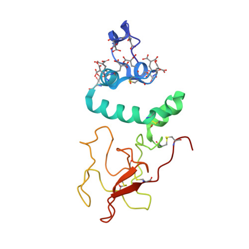
    1NL1, 1NL2

  • PubMed Abstract: 

    In a calcium-dependent interaction critical for blood coagulation, vitamin K-dependent blood coagulation proteins bind cell membranes containing phosphatidylserine via gamma-carboxyglutamic acid-rich (Gla) domains. Gla domain-mediated protein-membrane interaction is required for generation of thrombin, the terminal enzyme in the coagulation cascade, on a physiologic time scale. We determined by X-ray crystallography and NMR spectroscopy the lysophosphatidylserine-binding site in the bovine prothrombin Gla domain. The serine head group binds Gla domain-bound calcium ions and Gla residues 17 and 21, fixed elements of the Gla domain fold, predicting the structural basis for phosphatidylserine specificity among Gla domains. Gla domains provide a unique mechanism for protein-phospholipid membrane interaction. Increasingly Gla domains are being identified in proteins unrelated to blood coagulation. Thus, this membrane-binding mechanism may be important in other physiologic processes.


  • Organizational Affiliation
    • Center for Hemostasis and Thrombosis Research, Beth Israel Deaconess Medical Center and Department of Medicine, Harvard Medical School, 330 Brookline Avenue, Boston, Massachusetts 02215, USA.

Macromolecule Content 

  • Total Structure Weight: 18.38 kDa 
  • Atom Count: 1,323 
  • Modeled Residue Count: 146 
  • Deposited Residue Count: 146 
  • Unique protein chains: 1

Macromolecules

Find similar proteins by:|  3D Structure
Entity ID: 1
MoleculeChains  Sequence LengthOrganismDetailsImage
Prothrombin146Bos taurusMutation(s): 10 
EC: 3.4.21.5
Membrane Entity: Yes 
UniProt
Find proteins for P00735 (Bos taurus)
Explore P00735 
Go to UniProtKB:  P00735
Entity Groups
Sequence Clusters30% Identity50% Identity70% Identity90% Identity95% Identity100% Identity
UniProt GroupP00735
Glycosylation
Glycosylation Sites: 2
Sequence Annotations
Expand
Reference Sequence

Oligosaccharides

Help  
Entity ID: 2
MoleculeChains Length2D Diagram GlycosylationD Interactions
2-acetamido-2-deoxy-beta-D-glucopyranose-(1-4)-2-acetamido-2-deoxy-beta-D-glucopyranose
B
2N-Glycosylation
Glycosylation Resources
GlyTouCan: G42666HT
GlyCosmos: G42666HT
GlyGen: G42666HT

Small Molecules

Ligands 4 Unique
IDChains Name / Formula / InChI Key2D Diagram3D Interactions
LPS

Query on LPS


Download:Ideal Coordinates CCD File
N [auth A]O-{HYDROXY[((2R)-2-HYDROXY-3-{[(1S)-1-HYDROXYPENTADECYL]OXY}PROPYL)OXY]PHOSPHORYL}-L-SERINE
C21 H44 N O9 P
RPZLJDFLPRHXGM-HSALFYBXSA-N
NAG

Query on NAG


Download:Ideal Coordinates CCD File
C [auth A]2-acetamido-2-deoxy-beta-D-glucopyranose
C8 H15 N O6
OVRNDRQMDRJTHS-FMDGEEDCSA-N
CA

Query on CA


Download:Ideal Coordinates CCD File
D [auth A]
E [auth A]
F [auth A]
G [auth A]
H [auth A]
D [auth A],
E [auth A],
F [auth A],
G [auth A],
H [auth A],
I [auth A],
J [auth A]
CALCIUM ION
Ca
BHPQYMZQTOCNFJ-UHFFFAOYSA-N
CL

Query on CL


Download:Ideal Coordinates CCD File
K [auth A],
L [auth A],
M [auth A]
CHLORIDE ION
Cl
VEXZGXHMUGYJMC-UHFFFAOYSA-M
Modified Residues  1 Unique
IDChains TypeFormula2D DiagramParent
CGU
Query on CGU
A
L-PEPTIDE LINKINGC6 H9 N O6GLU

Experimental Data & Validation

Experimental Data

  • Method: X-RAY DIFFRACTION
  • Resolution: 2.30 Å
  • R-Value Free:  0.274 (Depositor) 
  • R-Value Work:  0.236 (Depositor) 
  • R-Value Observed: 0.236 (Depositor) 
Space Group: P 21 21 21
Unit Cell:
Length ( Å )Angle ( ˚ )
a = 38.894α = 90
b = 53.217β = 90
c = 128.959γ = 90
Software Package:
Software NamePurpose
CNSrefinement
DENZOdata reduction
SCALEPACKdata scaling
AMoREphasing

Structure Validation

View Full Validation Report


Entry History 

Deposition Data

Revision History  (Full details and data files)

  • Version 1.0: 2003-09-16
    Type: Initial release
  • Version 1.1: 2008-04-29
    Changes: Version format compliance
  • Version 1.2: 2011-07-13
    Changes: Non-polymer description, Version format compliance
  • Version 2.0: 2020-07-29
    Type: Remediation
    Reason: Carbohydrate remediation
    Changes: Advisory, Atomic model, Data collection, Database references, Derived calculations, Structure summary
  • Version 2.1: 2023-08-16
    Changes: Data collection, Database references, Refinement description, Structure summary
  • Version 2.2: 2023-11-15
    Changes: Data collection