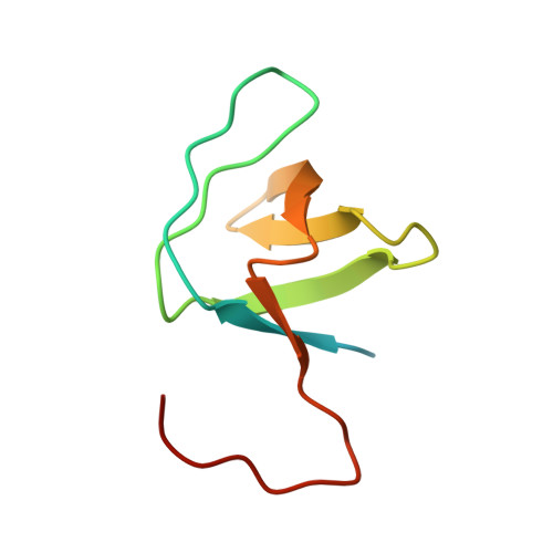
1NEG

Crystal Structure Analysis of N-and C-terminal labeled SH3-domain of alpha-Chicken Spectrin


Experimental Data Snapshot

  • Method: X-RAY DIFFRACTION
  • Resolution: 2.30 Å
  • R-Value Free: 0.249 
  • R-Value Work: 0.200 
  • R-Value Observed: 0.200 

Starting Model: experimental
View more details

wwPDB Validation   3D Report Full Report


This is version 1.5 of the entry. See complete history


Literature

Rapid purification and crystal structure analysis of a small protein carrying two terminal affinity tags

Mueller, U.Buessow, K.Diehl, A.Bartl, F.J.Niesen, F.H.Nyarsik, L.Heinemann, U.

(2003) J Struct Funct Genomics 4: 217-225

  • DOI: https://doi.org/10.1023/b:jsfg.0000016119.50040.a3
  • Primary Citation of Related Structures:  
    1NEG

  • PubMed Abstract: 

    Small peptide tags are often fused to proteins to allow their affinity purification in high-throughput structure analysis schemes. To assess the compatibility of small peptide tags with protein crystallization and to examine if the tags alter the three-dimensional structure, the N-terminus of the chicken alpha-spectrin SH3 domain was labeled with a His6 tag and the C-terminus with a StrepII tag. The resulting protein, His6-SH3-StrepII, consists of 83 amino-acid residues, 23 of which originate from the tags. His6-SH3-StrepII is readily purified by dual affinity chromatography, has very similar biophysical characteristics as the untagged protein domain and crystallizes readily from a number of sparse-matrix screen conditions. The crystal structure analysis at 2.3 A resolution proves native-like structure of His6-SH3-StrepII and shows the entire His6 tag and part of the StrepII tag to be disordered in the crystal. Obviously, the fused affinity tags did not interfere with crystallization and structure analysis and did not change the protein structure. From the extreme case of His6-SH3-StrepII, where affinity tags represent 27% of the total fusion protein mass, we extrapolate that protein constructs with N- and C-terminal peptide tags may lend themselves to biophysical and structural investigations in high-throughput regimes.


  • Organizational Affiliation

    Institut für Chemie/Kristallographie, Freie Universität Berlin, Takustr. 6, D-14195 Berlin, Germany.


Macromolecules
Find similar proteins by:  (by identity cutoff)  |  3D Structure
Entity ID: 1
MoleculeChains Sequence LengthOrganismDetailsImage
Spectrin alpha chain, brain83Gallus gallusMutation(s): 0 
UniProt
Find proteins for P07751 (Gallus gallus)
Explore P07751 
Go to UniProtKB:  P07751
Entity Groups  
Sequence Clusters30% Identity50% Identity70% Identity90% Identity95% Identity100% Identity
UniProt GroupP07751
Sequence Annotations
Expand
  • Reference Sequence
Small Molecules
Ligands 1 Unique
IDChains Name / Formula / InChI Key2D Diagram3D Interactions
AZI
Query on AZI

Download Ideal Coordinates CCD File 
B [auth A]AZIDE ION
N3
IVRMZWNICZWHMI-UHFFFAOYSA-N
Experimental Data & Validation

Experimental Data

  • Method: X-RAY DIFFRACTION
  • Resolution: 2.30 Å
  • R-Value Free: 0.249 
  • R-Value Work: 0.200 
  • R-Value Observed: 0.200 
  • Space Group: P 43 21 2
Unit Cell:
Length ( Å )Angle ( ˚ )
a = 41.865α = 90
b = 41.865β = 90
c = 92.865γ = 90
Software Package:
Software NamePurpose
DENZOdata reduction
SCALEPACKdata scaling
AMoREphasing
CNSrefinement

Structure Validation

View Full Validation Report


Entry History 

Deposition Data

Revision History  (Full details and data files)

  • Version 1.0: 2003-01-14
    Type: Initial release
  • Version 1.1: 2008-04-28
    Changes: Version format compliance
  • Version 1.2: 2011-07-13
    Changes: Version format compliance
  • Version 1.3: 2018-04-04
    Changes: Data collection
  • Version 1.4: 2022-12-21
    Changes: Database references, Derived calculations
  • Version 1.5: 2023-09-20
    Changes: Data collection, Refinement description