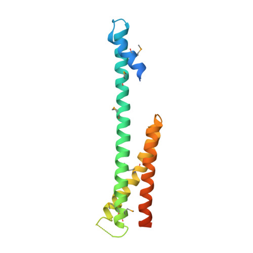
1NAF

Crystal structure of the human GGA1 GAT domain


Experimental Data Snapshot

  • Method: X-RAY DIFFRACTION
  • Resolution: 2.80 Å
  • R-Value Free: 0.283 
  • R-Value Work: 0.248 
  • R-Value Observed: 0.248 

wwPDB Validation   3D Report Full Report


This is version 1.3 of the entry. See complete history


Literature

The Structure of the GGA1-GAT Domain Reveals the Molecular Basis for ARF Binding and Membrane Association of GGAs

Collins, B.M.Watson, P.J.Owen, D.J.

(2003) Dev Cell 4: 321-332

  • DOI: https://doi.org/10.1016/s1534-5807(03)00037-6
  • Primary Citation of Related Structures:  
    1NAF

  • PubMed Abstract: 
  • The GGAs are a family of clathrin adaptor proteins involved in vesicular transport between the trans-Golgi network and endosomal system. Here we confirm reports that GGAs are targeted to the Golgi via interaction between the GGA-GAT domain and ARF-GTP, and we present the structure of the GAT domain of human GGA1, completing the structural description of the folded domains of GGA proteins ...

    The GGAs are a family of clathrin adaptor proteins involved in vesicular transport between the trans-Golgi network and endosomal system. Here we confirm reports that GGAs are targeted to the Golgi via interaction between the GGA-GAT domain and ARF-GTP, and we present the structure of the GAT domain of human GGA1, completing the structural description of the folded domains of GGA proteins. The GGA-GAT domain possesses an all alpha-helical fold with a "paper clip" topology comprising two independent subdomains. Structure-based mutagenesis demonstrates that ARF1-GTP binding by GGAs is exclusively governed by the N-terminal "hook" subdomain, and, using an in vitro recruitment assay, we show that ARF-GTP binding by this small structure is required and sufficient for Golgi targeting of GGAs.


    Organizational Affiliation

    Department of Clinical Biochemistry, University of Cambridge, Hills Road, CB2 2XY, Cambridge, United Kingdom. bmc25@cam.ac.uk


Macromolecules
Find similar proteins by:  (by identity cutoff)  |  3D Structure
Entity ID: 1
MoleculeChainsSequence LengthOrganismDetailsImage
ADP-ribosylation factor binding protein GGA1158Homo sapiensMutation(s): 0 
Gene Names: GGA1
UniProt & NIH Common Fund Data Resources
Find proteins for Q9UJY5 (Homo sapiens)
Explore Q9UJY5 
Go to UniProtKB:  Q9UJY5
PHAROS:  Q9UJY5
Entity Groups  
Sequence Clusters30% Identity50% Identity70% Identity90% Identity95% Identity100% Identity
UniProt GroupQ9UJY5
Protein Feature View
Expand
  • Reference Sequence
Small Molecules
Modified Residues  1 Unique
IDChainsTypeFormula2D DiagramParent
MSE
Query on MSE
A
L-PEPTIDE LINKINGC5 H11 N O2 SeMET
Experimental Data & Validation

Experimental Data

  • Method: X-RAY DIFFRACTION
  • Resolution: 2.80 Å
  • R-Value Free: 0.283 
  • R-Value Work: 0.248 
  • R-Value Observed: 0.248 
  • Space Group: P 63
Unit Cell:
Length ( Å )Angle ( ˚ )
a = 82.7α = 90
b = 82.7β = 90
c = 69.9γ = 120
Software Package:
Software NamePurpose
MOSFLMdata reduction
SCALAdata scaling
SHARPphasing
CNSrefinement
CCP4data scaling

Structure Validation

View Full Validation Report


Entry History 

Deposition Data

Revision History  (Full details and data files)

  • Version 1.0: 2003-03-25
    Type: Initial release
  • Version 1.1: 2008-04-28
    Changes: Version format compliance
  • Version 1.2: 2011-07-13
    Changes: Version format compliance
  • Version 1.3: 2018-02-14
    Changes: Experimental preparation, Structure summary