DNA recognition by the Anthracycline Antibiotic Respinomycin D: NMR Structure of the Intercalation Complex with d(AGACGTCT)2
Searle, M.S., Maynard, A.J., Williams, H.E.L.(2003) Org Biomol Chem 1: 60-66
- PubMed: 12929391 Search on PubMed
- DOI: https://doi.org/10.1039/b208622k
- Primary Citation Related Structures:
1N37 - PubMed Abstract:
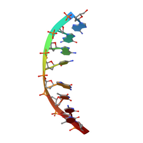
Respinomycin D is a member of the anthracycline family of antitumour antibiotics that interact with double stranded DNA through intercalation. The clinical agents daunomycin and doxorubicin are the most well-studied of this class but have a relatively simple molecular architecture in which the pendant daunosamine sugar resides in the DNA minor groove. Respinomycin D, which belongs to the nogalamycin group of anthracyclines, possesses additional sugar residues at either end of the aglycone chromophore that modulate the biological activity but whose role in molecular recognition is unknown. We report the NMR structure of the respinomycin D-d(AGACGTCT)2 complex in solution derived from NOE restraints and molecular dynamics simulations. We show that the drug threads through the DNA double helix forming stabilising interactions in both the major and minor groove, the latter through a different binding geometry to that previously reported. The bicycloaminoglucose sugar resides in the major groove and makes specific contacts with guanine at the 5'-CpG intercalation site, however, the disaccharide attached at the C4 position plays little part in drug binding and DNA recognition and is largely solvent exposed.
- School of Chemistry, University Park, University of Nottingham, Nottingham, UK NG7 2RD.
Organizational Affiliation: