1N0V

Crystal structure of elongation factor 2


Experimental Data Snapshot

  • Method: X-RAY DIFFRACTION
  • Resolution: 2.85 Å
  • R-Value Free: 0.273 
  • R-Value Work: 0.232 
  • R-Value Observed: 0.233 

wwPDB Validation   3D Report Full Report


This is version 1.4 of the entry. See complete history


Literature

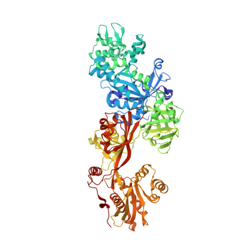
Two crystal structures demonstrate large conformational changes in the eukaryotic ribosomal translocase.

Joergensen, R.Ortiz, P.A.Carr-Schmid, A.Nissen, P.Kinzy, T.G.Andersen, G.R.

(2003) Nat Struct Biol 10: 379-385

  • DOI: https://doi.org/10.1038/nsb923
  • Primary Citation of Related Structures:  
    1N0U, 1N0V

  • PubMed Abstract: 

    Two crystal structures of yeast translation elongation factor 2 (eEF2) were determined: the apo form at 2.9 A resolution and eEF2 in the presence of the translocation inhibitor sordarin at 2.1 A resolution. The overall conformation of apo eEF2 is similar to that of its prokaryotic homolog elongation factor G (EF-G) in complex with GDP. Upon sordarin binding, the three tRNA-mimicking C-terminal domains undergo substantial conformational changes, while the three N-terminal domains containing the nucleotide-binding site form an almost rigid unit. The conformation of eEF2 in complex with sordarin is entirely different from known conformations observed in crystal structures of EF-G or from cryo-EM studies of EF-G-70S complexes. The domain rearrangements induced by sordarin binding and the highly ordered drug-binding site observed in the eEF2-sordarin structure provide a high-resolution structural basis for the mechanism of sordarin inhibition. The two structures also emphasize the dynamic nature of the ribosomal translocase.


  • Organizational Affiliation

    Department of Molecular Biology, Aarhus University, Gustav Wieds vej 10C, DK8000 Arhus, Denmark.


Macromolecules
Find similar proteins by:  (by identity cutoff)  |  3D Structure
Entity ID: 1
MoleculeChains Sequence LengthOrganismDetailsImage
Elongation factor 2A [auth C],
B [auth D]
842Saccharomyces cerevisiaeMutation(s): 0 
UniProt
Find proteins for P32324 (Saccharomyces cerevisiae (strain ATCC 204508 / S288c))
Explore P32324 
Go to UniProtKB:  P32324
Entity Groups  
Sequence Clusters30% Identity50% Identity70% Identity90% Identity95% Identity100% Identity
UniProt GroupP32324
Sequence Annotations
Expand
  • Reference Sequence
Experimental Data & Validation

Experimental Data

  • Method: X-RAY DIFFRACTION
  • Resolution: 2.85 Å
  • R-Value Free: 0.273 
  • R-Value Work: 0.232 
  • R-Value Observed: 0.233 
  • Space Group: P 21 21 21
Unit Cell:
Length ( Å )Angle ( ˚ )
a = 98.835α = 90
b = 114.546β = 90
c = 177.31γ = 90
Software Package:
Software NamePurpose
DENZOdata reduction
SCALEPACKdata scaling
AMoREphasing
CNSrefinement

Structure Validation

View Full Validation Report


Entry History 

Deposition Data

Revision History  (Full details and data files)

  • Version 1.0: 2002-11-27
    Type: Initial release
  • Version 1.1: 2008-04-28
    Changes: Version format compliance
  • Version 1.2: 2011-07-13
    Changes: Version format compliance
  • Version 1.3: 2018-01-31
    Changes: Data collection, Experimental preparation
  • Version 1.4: 2024-02-14
    Changes: Data collection, Database references