Probing the function, conformational plasticity, and dimer-dimer contacts of the GluR2 ligand-binding core: studies of 5-substituted willardiines and GluR2 S1S2 in the crystal.
Jin, R., Gouaux, E.(2003) Biochemistry 42: 5201-5213
- PubMed: 12731861 Search on PubMed
- DOI: https://doi.org/10.1021/bi020632t
- Primary Citation Related Structures:
1MXU, 1MXV, 1MXW, 1MXX, 1MXY, 1MXZ, 1MY0, 1MY1, 1MY2, 1MY3, 1MY4 - PubMed Abstract:
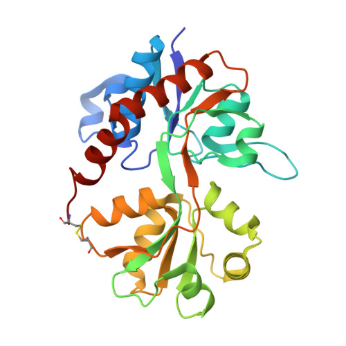
Numerous naturally occurring and synthetic alpha-amino acids act as agonists on (S)-2-amino-3-(3-hydroxy-5-methyl-4-isoxazole) propionic acid (AMPA) receptors but nevertheless display significant differences in their functional properties and modes of interaction. The 5-substituted willardiines are a series of compounds that exhibit a range of affinities, act as partial agonists, and give rise to intermediate levels of activation and desensitization. However, the molecular basis for the activities of 5-substituted willardiines has not been conclusively elaborated at the level of atomic resolution. Here we provide insight into the molecular basis of the potency and efficacy elicited by the 5-substituted willardiines on the basis of cocrystal structures with the GluR2 ligand-binding core. We also show that the crystallized ligand-binding core has an affinity for agonists similar to the ligand-binding core in solution. Analysis of multiple crystal lattices suggests modes by which the ligand-binding core dimers interact in the tetrameric receptor. These studies further our understanding of how subtle differences in the structures of agonists are correlated to changes in the conformation of residues and water molecules in the immediate binding pocket and to the degree of domain closure.
- Department of Biochemistry and Molecular Biophysics, Columbia University, New York, New York 10032, USA.
Organizational Affiliation: