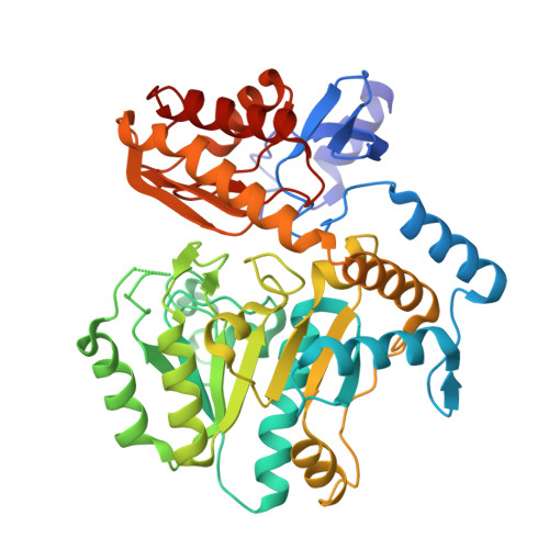
1MLZ

Crystal Structure of 7,8-Diaminopelargonic Acid Synthase in complex with the trans-isomer of amiclenomycin.


Experimental Data Snapshot

  • Method: X-RAY DIFFRACTION
  • Resolution: 2.15 Å
  • R-Value Free: 0.240 
  • R-Value Work: 0.205 
  • R-Value Observed: 0.207 

wwPDB Validation   3D Report Full Report


Ligand Structure Quality Assessment 


This is version 1.4 of the entry. See complete history


Literature

Structural basis for the inhibition of the biosynthesis of biotin by the antibiotic amiclenomycin

Sandmark, J.Mann, S.Marquet, A.Schneider, G.

(2002) J Biol Chem 277: 43352-43358

  • DOI: https://doi.org/10.1074/jbc.M207239200
  • Primary Citation of Related Structures:  
    1MLY, 1MLZ

  • PubMed Abstract: 

    The antibiotic amiclenomycin blocks the biosynthesis of biotin by inhibiting the pyridoxal-phosphate-dependent enzyme diaminopelargonic acid synthase. Inactivation of the enzyme is stereoselective, i.e. the cis isomer of amiclenomycin is a potent inhibitor, whereas the trans isomer is much less reactive. The crystal structure of the complex of the holoenzyme and amiclenomycin at 1.8 A resolution reveals that the internal aldimine linkage between the cofactor and the side chain of the catalytic residue Lys-274 is broken. Instead, a covalent bond is formed between the 4-amino nitrogen of amiclenomycin and the C4' carbon atom of pyridoxal-phosphate. The electron density for the bound inhibitor suggests that aromatization of the cyclohexadiene ring has occurred upon formation of the covalent adduct. This process could be initiated by proton abstraction at the C4 carbon atom of the cyclohexadiene ring, possibly by the proximal side chain of Lys-274, leading to the tautomer Schiff base followed by the removal of the second allylic hydrogen. The carboxyl tail of the amiclenomycin moiety forms a salt link to the conserved residue Arg-391 in the substrate-binding site. Modeling suggests steric hindrance at the active site as the determinant of the weak inhibiting potency of the trans isomer.


  • Organizational Affiliation

    Department of Medical Biochemistry and Biophysics, Karolinska Institutet, S-171 77 Stockholm, Sweden.


Macromolecules
Find similar proteins by:  (by identity cutoff)  |  3D Structure
Entity ID: 1
MoleculeChains Sequence LengthOrganismDetailsImage
7,8-diamino-pelargonic acid aminotransferase
A, B
429Escherichia coliMutation(s): 0 
Gene Names: bioA
EC: 2.6.1.62
UniProt
Find proteins for P12995 (Escherichia coli (strain K12))
Explore P12995 
Go to UniProtKB:  P12995
Entity Groups  
Sequence Clusters30% Identity50% Identity70% Identity90% Identity95% Identity100% Identity
UniProt GroupP12995
Sequence Annotations
Expand
  • Reference Sequence
Experimental Data & Validation

Experimental Data

  • Method: X-RAY DIFFRACTION
  • Resolution: 2.15 Å
  • R-Value Free: 0.240 
  • R-Value Work: 0.205 
  • R-Value Observed: 0.207 
  • Space Group: C 1 2 1
Unit Cell:
Length ( Å )Angle ( ˚ )
a = 128.034α = 90
b = 55.96β = 109.65
c = 116.001γ = 90
Software Package:
Software NamePurpose
MOSFLMdata reduction
SCALAdata scaling
AMoREphasing
REFMACrefinement
CCP4data scaling

Structure Validation

View Full Validation Report


Ligand Structure Quality Assessment 


Entry History 

Deposition Data

Revision History  (Full details and data files)

  • Version 1.0: 2002-12-04
    Type: Initial release
  • Version 1.1: 2008-04-28
    Changes: Version format compliance
  • Version 1.2: 2011-07-13
    Changes: Non-polymer description, Version format compliance
  • Version 1.3: 2018-03-07
    Changes: Advisory, Data collection
  • Version 1.4: 2024-02-14
    Changes: Advisory, Data collection, Database references, Derived calculations