1MIJ | pdb_00001mij

Crystal Structure of the Homeo-prospero Domain of D. melanogaster Prospero


Experimental Data Snapshot

  • Method: X-RAY DIFFRACTION
  • Resolution: 2.05 Å
  • R-Value Free: 
    0.256 (Depositor), 0.220 (DCC) 
  • R-Value Work: 
    0.216 (Depositor), 0.213 (DCC) 

wwPDB Validation 3D Report Full Report

Validation slider image for 1MIJ

This is version 1.3 of the entry. See complete history

Literature

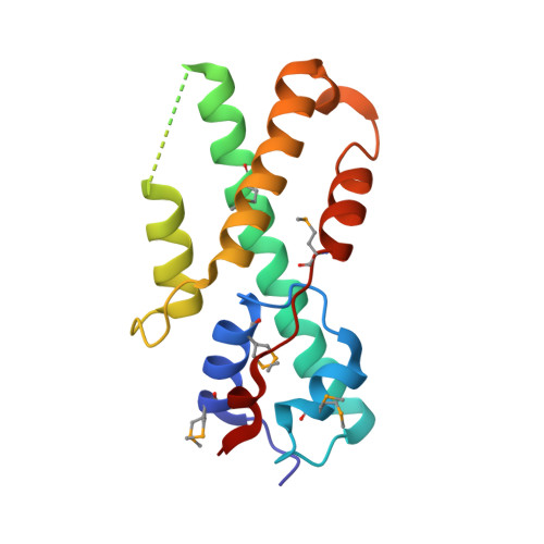
Structure of the DNA Binding Region of Prospero Reveals a Novel Homeo-Prospero Domain

Ryter, J.M.Doe, C.Q.Matthews, B.W.

(2002) Structure 10: 1541-1549

  • DOI: https://doi.org/10.1016/s0969-2126(02)00883-3
  • Primary Citation Related Structures: 
    1MIJ

  • PubMed Abstract: 

    The Prospero transcription factor promotes neural differentiation in Drosophila, and its activity is tightly regulated by modulating its subcellular localization. Prospero is exported from the nucleus of neural precursors but imported into the nucleus of daughter cells, which is necessary for their proper differentiation. Prospero has a highly divergent putative homeodomain adjacent to a conserved Prospero domain; both are required for sequence-specific DNA binding. Here we show that the structure of these two regions consists of a single structural unit (a homeo-prospero domain), in which the Prospero domain region is in position to contribute to DNA binding and also to mask a defined nuclear export signal that is within the putative homeodomain region. We propose that the homeo-prospero domain coordinately regulates Prospero nuclear localization and DNA binding specificity.


  • Organizational Affiliation
    • Institute of Molecular Biology, Howard Hughes Medical Institute and Department of Physics, 1229 University of Oregon, Eugene, OR 97403, USA.

Macromolecule Content 

  • Total Structure Weight: 18.45 kDa 
  • Atom Count: 1,434 
  • Modeled Residue Count: 139 
  • Deposited Residue Count: 152 
  • Unique protein chains: 1

Macromolecules

Find similar proteins by:|  3D Structure
Entity ID: 1
MoleculeChains  Sequence LengthOrganismDetailsImage
Protein prospero152Drosophila melanogasterMutation(s): 5 
Gene Names: PROS
UniProt
Find proteins for P29617 (Drosophila melanogaster)
Explore P29617 
Go to UniProtKB:  P29617
Entity Groups
Sequence Clusters30% Identity50% Identity70% Identity90% Identity95% Identity100% Identity
UniProt GroupP29617
Sequence Annotations
Expand
Reference Sequence

Small Molecules

Modified Residues  1 Unique
IDChains TypeFormula2D DiagramParent
MSE
Query on MSE
A
L-PEPTIDE LINKINGC5 H11 N O2 SeMET

Experimental Data & Validation

Experimental Data

  • Method: X-RAY DIFFRACTION
  • Resolution: 2.05 Å
  • R-Value Free:  0.256 (Depositor), 0.220 (DCC) 
  • R-Value Work:  0.216 (Depositor), 0.213 (DCC) 
Space Group: P 1 21 1
Unit Cell:
Length ( Å )Angle ( ˚ )
a = 35.3α = 90
b = 49.9β = 97.4
c = 51.5γ = 90
Software Package:
Software NamePurpose
DENZOdata reduction
SCALEPACKdata scaling
SHARPphasing
CNSrefinement

Structure Validation

View Full Validation Report


Entry History 

Deposition Data

Revision History  (Full details and data files)

  • Version 1.0: 2002-12-04
    Type: Initial release
  • Version 1.1: 2008-04-28
    Changes: Version format compliance
  • Version 1.2: 2011-07-13
    Changes: Version format compliance
  • Version 1.3: 2024-11-06
    Changes: Data collection, Database references, Derived calculations, Structure summary