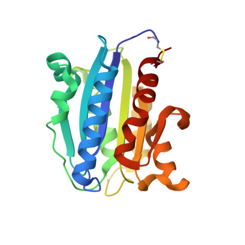
Engineered allosteric mutants of the integrin alphaMbeta2 I domain: structural and functional studies
McCleverty, C.J., Liddington, R.C.(2003) Biochem J 372: 121-127
- PubMed: 12611591
- DOI: https://doi.org/10.1042/BJ20021273
- Primary Citation of Related Structures:
1MF7, 1N9Z, 1NA5 - PubMed Abstract:
The alpha-I domain, found in the alpha-subunit of the leucocyte integrins such as alphaMbeta2 and alphaLbeta2, switches between the open and closed tertiary conformations, reflecting the high- and low-affinity ligand-binding states of the integrin that are required for regulated cell adhesion and migration. In the present study we show, by using point mutations and engineered disulphide bonds, that ligand affinity can be reduced or increased allosterically by altering the equilibrium between the closed and open states. We determined equilibrium constants for the binding of two ligands, fibrinogen and intercellular cell-adhesion molecule 1, to the alphaM-I domain by surface plasmon resonance, and determined crystal structures of a low-affinity mutant. Locking the domain in the open conformation increases affinity by a factor of no greater than 10, consistent with a closely balanced equilibrium between the two conformations in the absence of ligand. This behaviour contrasts with that of the unliganded alphaL-I domain, for which the equilibrium lies strongly in favour of the closed conformation. These results suggest significant differences in the way the parent integrins regulate I domain conformation and hence ligand affinity.
- The Burnham Institute, 10901 North Torrey Pines Rd, La Jolla, CA 92037, U.S.A.
Organizational Affiliation:


