1N9Z | pdb_00001n9z

INTEGRIN ALPHA M I DOMAIN MUTANT


Experimental Data Snapshot

  • Method: X-RAY DIFFRACTION
  • Resolution: 2.50 Å
  • R-Value Free: 
    0.278 (Depositor), 0.289 (DCC) 
  • R-Value Work: 
    0.242 (Depositor), 0.246 (DCC) 
  • R-Value Observed: 
    0.247 (Depositor) 

Starting Model: experimental
View more details

wwPDB Validation 3D Report Full Report

Validation slider image for 1N9Z

This is version 1.4 of the entry. See complete history

Literature

Engineered allosteric mutants of the integrin alphaMbeta2 I domain: structural and functional studies

McCleverty, C.J.Liddington, R.C.

(2003) Biochem J 372: 121-127

  • DOI: https://doi.org/10.1042/BJ20021273
  • Primary Citation Related Structures: 
    1MF7, 1N9Z, 1NA5

  • PubMed Abstract: 

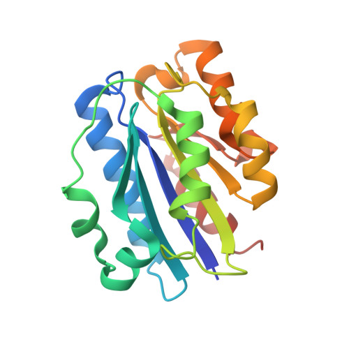
    The alpha-I domain, found in the alpha-subunit of the leucocyte integrins such as alphaMbeta2 and alphaLbeta2, switches between the open and closed tertiary conformations, reflecting the high- and low-affinity ligand-binding states of the integrin that are required for regulated cell adhesion and migration. In the present study we show, by using point mutations and engineered disulphide bonds, that ligand affinity can be reduced or increased allosterically by altering the equilibrium between the closed and open states. We determined equilibrium constants for the binding of two ligands, fibrinogen and intercellular cell-adhesion molecule 1, to the alphaM-I domain by surface plasmon resonance, and determined crystal structures of a low-affinity mutant. Locking the domain in the open conformation increases affinity by a factor of no greater than 10, consistent with a closely balanced equilibrium between the two conformations in the absence of ligand. This behaviour contrasts with that of the unliganded alphaL-I domain, for which the equilibrium lies strongly in favour of the closed conformation. These results suggest significant differences in the way the parent integrins regulate I domain conformation and hence ligand affinity.


  • Organizational Affiliation
    • The Burnham Institute, 10901 North Torrey Pines Rd, La Jolla, CA 92037, U.S.A.

Macromolecule Content 

  • Total Structure Weight: 22 kDa 
  • Atom Count: 1,486 
  • Modeled Residue Count: 184 
  • Deposited Residue Count: 192 
  • Unique protein chains: 1

Macromolecules

Find similar proteins by:|  3D Structure
Entity ID: 1
MoleculeChains  Sequence LengthOrganismDetailsImage
Integrin alpha-M192Homo sapiensMutation(s): 3 
Gene Names: ITGAM OR CR3A OR CD11B
UniProt & NIH Common Fund Data Resources
Find proteins for P11215 (Homo sapiens)
Explore P11215 
Go to UniProtKB:  P11215
PHAROS:  P11215
GTEx:  ENSG00000169896 
Entity Groups
Sequence Clusters30% Identity50% Identity70% Identity90% Identity95% Identity100% Identity
UniProt GroupP11215
Sequence Annotations
Expand
Reference Sequence

Small Molecules

Ligands 1 Unique
IDChains Name / Formula / InChI Key2D Diagram3D Interactions
MG

Query on MG


Download:Ideal Coordinates CCD File
B [auth A]MAGNESIUM ION
Mg
JLVVSXFLKOJNIY-UHFFFAOYSA-N

Experimental Data & Validation

Experimental Data

  • Method: X-RAY DIFFRACTION
  • Resolution: 2.50 Å
  • R-Value Free:  0.278 (Depositor), 0.289 (DCC) 
  • R-Value Work:  0.242 (Depositor), 0.246 (DCC) 
  • R-Value Observed: 0.247 (Depositor) 
Space Group: P 43
Unit Cell:
Length ( Å )Angle ( ˚ )
a = 44.22α = 90
b = 44.22β = 90
c = 99.91γ = 90
Software Package:
Software NamePurpose
DENZOdata reduction
SCALEPACKdata scaling
AMoREphasing
CNSrefinement

Structure Validation

View Full Validation Report


Entry History 

Deposition Data

Revision History  (Full details and data files)

  • Version 1.0: 2003-05-20
    Type: Initial release
  • Version 1.1: 2008-04-28
    Changes: Version format compliance
  • Version 1.2: 2011-07-13
    Changes: Version format compliance
  • Version 1.3: 2021-10-27
    Changes: Database references, Derived calculations
  • Version 1.4: 2023-08-16
    Changes: Data collection, Refinement description